. 2012;7(10):e46507.
doi: 10.1371/journal.pone.0046507. Epub 2012 Oct 8.

Initial characterization of the Pf-Int recombinase from the malaria parasite Plasmodium falciparum

Affiliations

Initial characterization of the Pf-Int recombinase from the malaria parasite Plasmodium falciparum

Mehdi Ghorbal et al. PLoS One. 2012.

Abstract

Background: Genetic variation is an essential means of evolution and adaptation in many organisms in response to environmental change. Certain DNA alterations can be carried out by site-specific recombinases (SSRs) that fall into two families: the serine and the tyrosine recombinases. SSRs are seldom found in eukaryotes. A gene homologous to a tyrosine site-specific recombinase has been identified in the genome of Plasmodium falciparum. The sequence is highly conserved among five other members of Plasmodia.

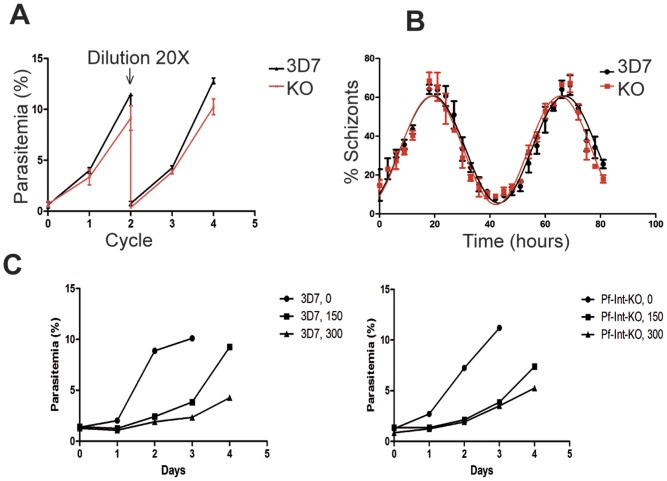
Methodology/principal findings: The predicted open reading frame encodes for a ∼57 kDa protein containing a C-terminal domain including the putative tyrosine recombinase conserved active site residues R-H-R-(H/W)-Y. The N-terminus has the typical alpha-helical bundle and potentially a mixed alpha-beta domain resembling that of λ-Int. Pf-Int mRNA is expressed differentially during the P. falciparum erythrocytic life stages, peaking in the schizont stage. Recombinant Pf-Int and affinity chromatography of DNA from genomic or synthetic origin were used to identify potential DNA targets after sequencing or micro-array hybridization. Interestingly, the sequences captured also included highly variable subtelomeric genes such as var, rif, and stevor sequences. Electrophoretic mobility shift assays with DNA were carried out to verify Pf-Int/DNA binding. Finally, Pf-Int knock-out parasites were created in order to investigate the biological role of Pf-Int.

Conclusions/significance: Our data identify for the first time a malaria parasite gene with structural and functional features of recombinases. Pf-Int may bind to and alter DNA, either in a sequence specific or in a non-specific fashion, and may contribute to programmed or random DNA rearrangements. Pf-Int is the first molecular player identified with a potential role in genome plasticity in this pathogen. Finally, Pf-Int knock-out parasite is viable showing no detectable impact on blood stage development, which is compatible with such function.


Conflict of interest statement

Competing Interests: The authors have declared that no competing interests exist.

Figures

Figure 1
Figure 1. Pf-Int is a tyrosine recombinase.
A) Schematic representation of domain organization of Pf-Int. The integrase is 490 amino acids long and is predicted to have two DNA-binding domains, the Arm binding domain (117–156 aa residues) and the core-binding domain (192–490 aa residues). The core binding domain is made of the two domains classically found in Y-SSR: the N-terminal domain (green) and the C-terminal catalytic domain (blue) that contains the catalytic residues R-K-(Y/H)-R-H/W-Y. B) Sequence alignment using Pf-Int and members of the tyrosine recombinase family (λ-integrase, Cre, XerD, XerC, IntI1, IntI4). Conserved catalytic residues (R-K-H-R-H/W) are depicted in green. The catalytic tyrosine is depicted in red.
Figure 2
Figure 2. Pf-Int is non-essential for the intra-erythrocytic parasite growth.
A) The effect of the gene disruption was examined by comparison of synchronized cultures of WT (black line) and KO (red line) parasites for four cycles. The cultures were diluted 20 times at the end of the second cycle. Values are the mean of two independent experiments whose standard deviation is shown by the bars. B) Synchronous cultures of the WT (black circles) and KO (red squares) parasites were grown for 80 hours and sampled every 3 h. The parasite stages were analyzed by flow cytometry and the percentage of parasites at the schizont stage was plotted against time. Values are the mean of two independent experiments whose standard deviation is shown by the bars. The data were fitted to an exponential sine wave equation using Prism program, and the fitting curves are shown. Black line corresponds to the WT cultures, and the red line to the KO cultures. C) Response to UV-induced DNA damage in 3D7 and Pf-Int-KO parasites. The effect of the gene disruption was examined by comparison of WT 3D7 (Left panel) and Pf-Int-KO (Right panel) parasites after UV irradiation at different doses (0, 150 and 300×100 µJ/cm2). Synchronous cultures were grown for 4 days after irradiation and sampled every day. The parasitemia was determined by flow cytometry. One representative experiment of two is shown.
Figure 3
Figure 3. Circular dichroism analysis of the purified recombinant proteins Pf-Int-C162 and Pf-Int-C192.
A) CD analysis in far UV. Black dots: experimental CD data. Blue line: modeled CD spectra of Pf-Int-C192. Red line: modeled CD spectra of Pf-Int-C162. The CD spectral analysis showed for both analyzed forms a high degree of α-helical organization and a low percentage of β-sheet (54/60% α-helical and 6/3% β-sheet for Pf-Int-C162/C192). B, C) Stability analysis by thermal denaturation coupled to CD for B) Pf-Int-C162 and C) Pf-Int-C192. Thermal CD transition curves (red lines) for Pf-Int-C162 and Pf-Int-C192 showed a beginning of stability change at approximately 50°C and 40°C, with a completion at 60°C and 50°C. The increase of the dynode signal (blue lines) indicates that the proteins are precipitating at the transition and thus the melting temperatures cannot be precisely determined.
Figure 4
Figure 4. Identified DNA targets of Pf-Int.
A) Clustal sequence alignment of the targets retained by SELEX method showed that the motif: (N)6CAANC(A/C)(N)2GT(N)3CG(N)7C(N)4 seems to be present in all of them. Conserved positions are shown by (*). B) Distribution of gDNA fragments retained by Pf-Int from genomic DNA capture. Y-axis: number of occurrences. X-axis: identity. Single hits are not labeled, but present in Table S2.
Figure 5
Figure 5. Characterization of Pf-Int interaction with DNA targets.
10 nM of labeled Selex8-DNA were incubated with increasing amounts of Pf-Int-C192 (lanes 1 to 6) and Pf-Int-C162 (lanes 7 to 12) A) in absence of poly (dI-dC) and B) in the presence of 10 µg/ml of poly (dI-dC).

References

    1. WHO (2010) Malaria Fact Sheet.
    1. Scherf A, Lopez-Rubio JJ, Riviere L (2008) Antigenic variation in Plasmodium falciparum. Annu Rev Microbiol 62: 445–470. - PubMed
    1. Deitsch KW, Lukehart SA, Stringer JR (2009) Common strategies for antigenic variation by bacterial, fungal and protozoan pathogens. Nat Rev Microbiol 7: 493–503. - PMC - PubMed
    1. Freitas-Junior LH, Bottius E, Pirrit LA, Deitsch KW, Scheidig C, et al. (2000) Frequent ectopic recombination of virulence factor genes in telomeric chromosome clusters of P. falciparum. Nature 407: 1018–1022. - PubMed
    1. Chaconas G, Kobryn K (2010) Structure, function, and evolution of linear replicons in Borrelia. Annu Rev Microbiol 64: 185–202. - PubMed

Publication types