. 2012;7(10):e46609.
doi: 10.1371/journal.pone.0046609. Epub 2012 Oct 4.

Cell stiffness is a biomarker of the metastatic potential of ovarian cancer cells

Affiliations

Cell stiffness is a biomarker of the metastatic potential of ovarian cancer cells

Wenwei Xu et al. PLoS One. 2012.

Abstract

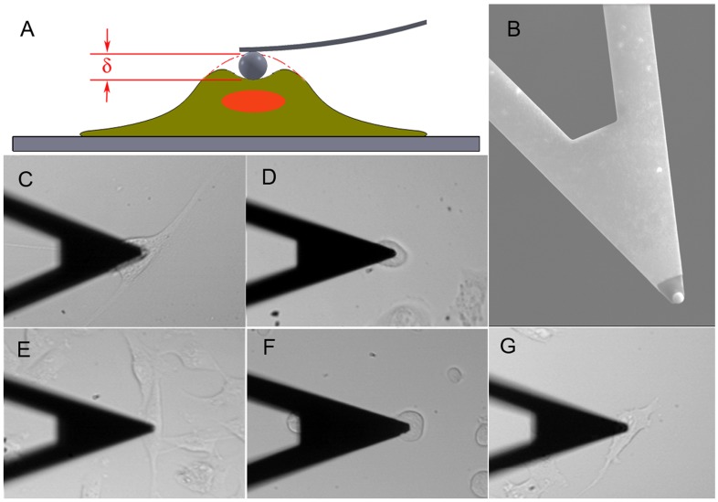
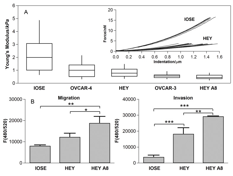
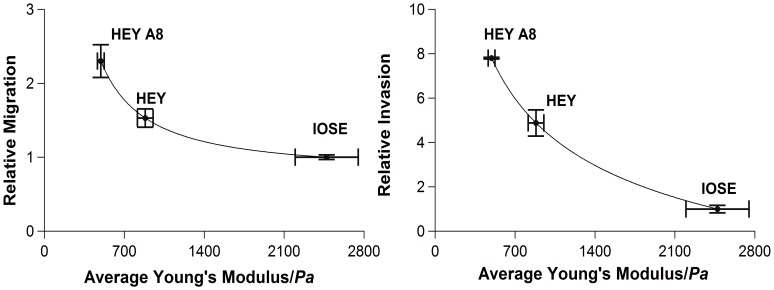
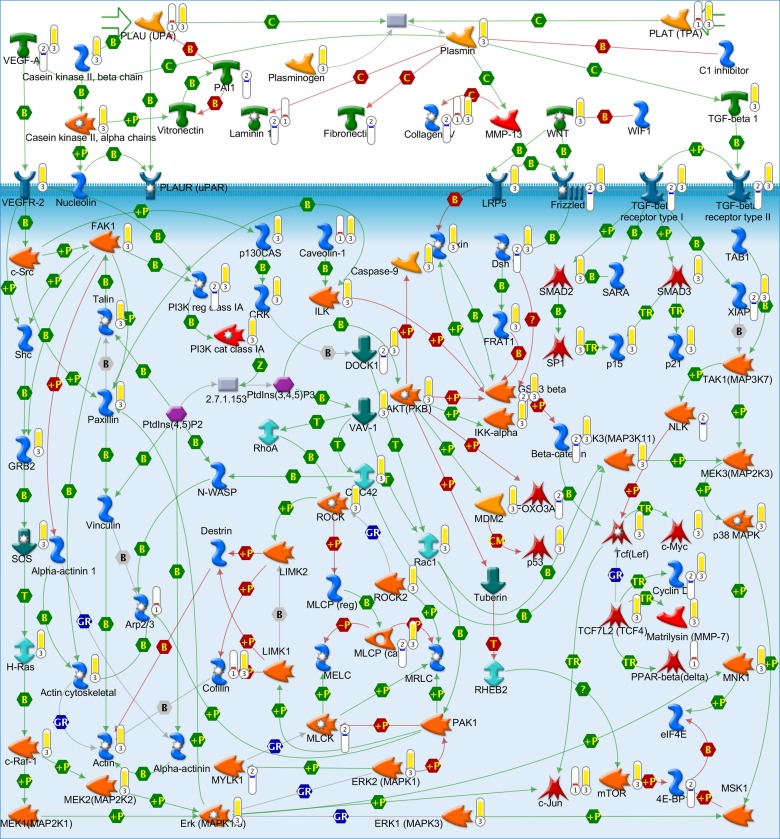
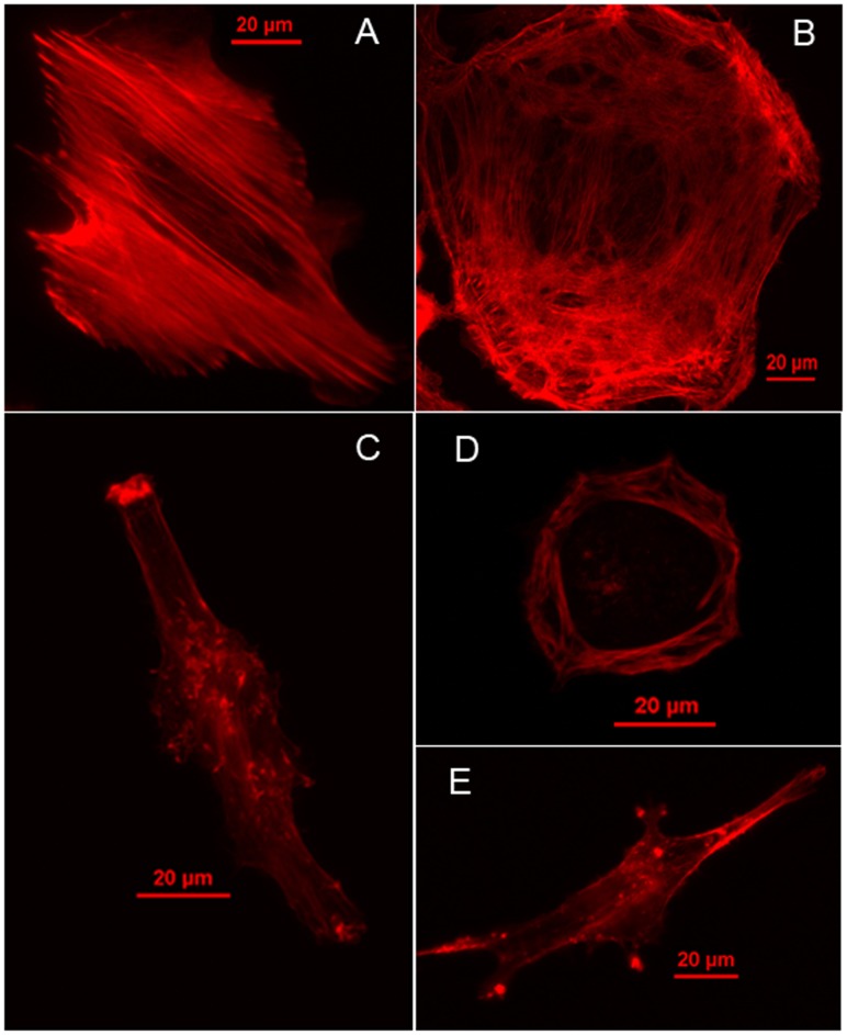
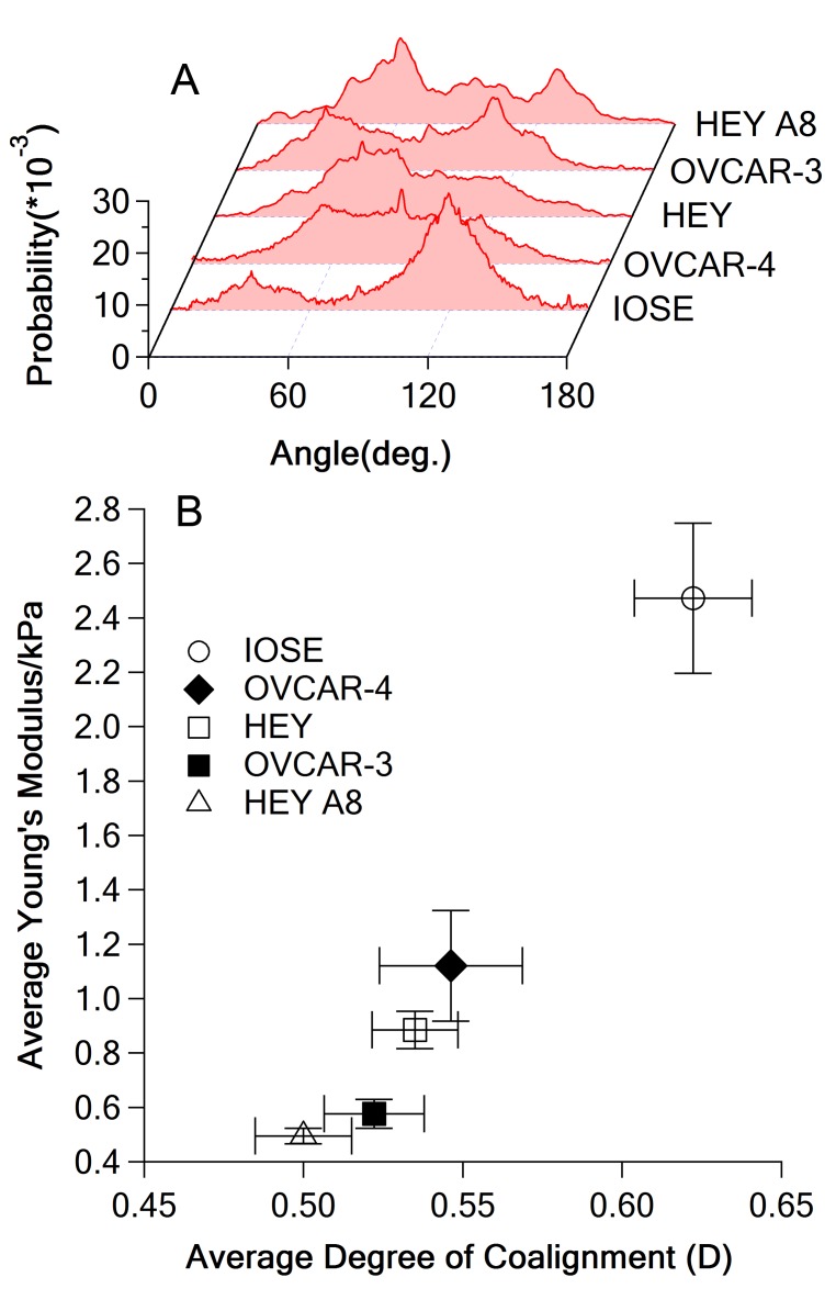
The metastatic potential of cells is an important parameter in the design of optimal strategies for the personalized treatment of cancer. Using atomic force microscopy (AFM), we show, consistent with previous studies conducted in other types of epithelial cancer, that ovarian cancer cells are generally softer and display lower intrinsic variability in cell stiffness than non-malignant ovarian epithelial cells. A detailed examination of highly invasive ovarian cancer cells (HEY A8) relative to their less invasive parental cells (HEY), demonstrates that deformability is also an accurate biomarker of metastatic potential. Comparative gene expression analyses indicate that the reduced stiffness of highly metastatic HEY A8 cells is associated with actin cytoskeleton remodeling and microscopic examination of actin fiber structure in these cell lines is consistent with this prediction. Our results indicate that cell stiffness may be a useful biomarker to evaluate the relative metastatic potential of ovarian and perhaps other types of cancer cells.


Conflict of interest statement

Competing Interests: The authors have declared that no competing interests exist.

Figures

Figure 1
Figure 1. Schematics of experiments.
(A) Sketch of measurements on cells with AFM, δ is indentation, (B) SEM image of the beaded tip, stiffness measurements of single cells with AFM for (C) IOSE, (D) OVCAR-4, (E) HEY, (F) OVCAR-3 and (G) HEY A8 cells. Same cantilever was used; the arm of the cantilever has a width of 20 µm and serves as a scale bar.
Figure 2
Figure 2. Stiffness distribution of cells and results of migration and invasion test.
(A) Box-and-whisker plots of stiffness of single cells for different cell lines, the percentiles are 10%, 25%, 50%, 75% and 90%, the inset shows the representative force curves of IOSE and HEY. Overall difference among means is significant (p-value<2.2×10⁻¹⁶, Kruskal-Wallis); pairwise differences are significant between IOSE and HEY, HEY A8 and OVCAR-3 cells, between HEY A8 and HEY cells and between HEY A8 and OVCAR-4 cells (p<0.05, Dunn’s post test); (B) Migration and invasion tests for IOSE, HEY and HEY A8 cells. F(480/520) is a fluorescence intensity at 480 nm excitation and 520 nm emission, which is proportional to the number of migrating or invading cells.
Figure 3
Figure 3. Scatterplots of relative migration and invasion versus average stiffness for IOSE, HEY and HEY A8 cells (migration and invasion of IOSE cells = 1).
The data points are fitted with power law for clarity. Error bars: standard errors of means.
Figure 4
Figure 4. TGF, WNT and cytoskeletal remodeling GeneGO pathway.
Red thermometer: genes transcriptionally up-regulated in HEY A8 cells; blue thermometer: genes transcriptionally down-regulated in HEY A8 cells; yellow thermometer: proteins topologically relevant to the set of up-regulated genes.
Figure 5
Figure 5. Fluorescence images of F-actin.
(A) IOSE, (B) OVCAR-4, (C) HEY, (D) OVCAR-3 and (E) HEY A8.
Figure 6
Figure 6. Relationship of F-actin coalignment and stiffness.
(A) Representative orientation distribution function for each cell line, (B) stiffness versus degree of coalignment of F-actin, error bar represents standard error of mean (SEM).

References

    1. Fletcher DA, Mullins RD (2010) Cell mechanics and the cytoskeleton. Nature 463: 485–492. - PMC - PubMed
    1. Bhadriraju K, Hansen LK (2002) Extracellular matrix- and cytoskeleton-dependent changes in cell shape and stiffness. Experimental Cell Research 278: 92–100. - PubMed
    1. Cross SE, Jin Y-S, Rao J, Gimzewski JK (2007) Nanomechanical analysis of cells from cancer patients. Nat Nano 2: 780–783. - PubMed
    1. Yamazaki D, Kurisu S, Takenawa T (2005) Regulation of cancer cell motility through actin reorganization. Cancer Science 96: 379–386. - PMC - PubMed
    1. Guck J, Schinkinger S, Lincoln B, Wottawah F, Ebert S, et al. (2005) Optical deformability as an inherent cell marker for testing malignant transformation and metastatic competence. Biophysical Journal 88: 3689–3698. - PMC - PubMed

Publication types