. 2012;7(10):e46623.
doi: 10.1371/journal.pone.0046623. Epub 2012 Oct 2.

The genome of Chelonid herpesvirus 5 harbors atypical genes

Affiliations

The genome of Chelonid herpesvirus 5 harbors atypical genes

Mathias Ackermann et al. PLoS One. 2012.

Abstract

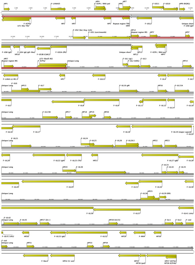
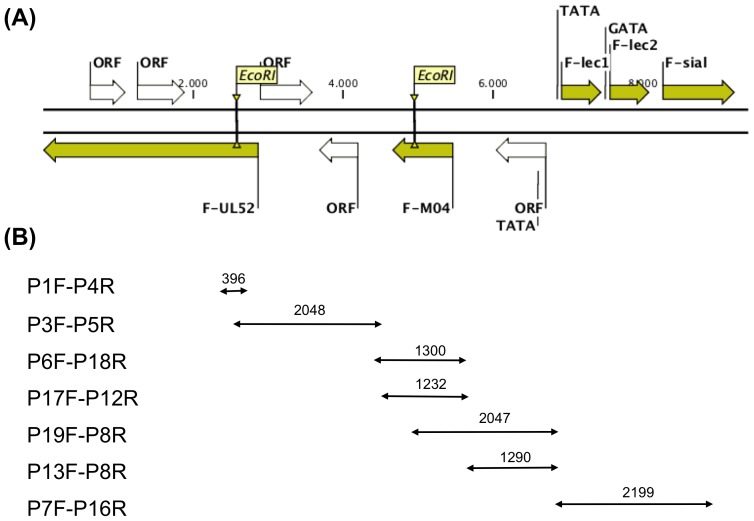
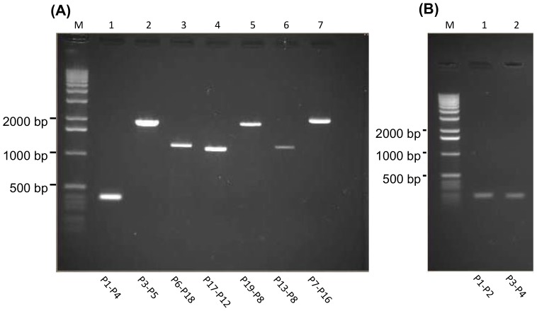
The Chelonid fibropapilloma-associated herpesvirus (CFPHV; ChHV5) is believed to be the causative agent of fibropapillomatosis (FP), a neoplastic disease of marine turtles. While clinical signs and pathology of FP are well known, research on ChHV5 has been impeded because no cell culture system for its propagation exists. We have cloned a BAC containing ChHV5 in pTARBAC2.1 and determined its nucleotide sequence. Accordingly, ChHV5 has a type D genome and its predominant gene order is typical for the varicellovirus genus within the alphaherpesvirinae. However, at least four genes that are atypical for an alphaherpesvirus genome were also detected, i.e. two members of the C-type lectin-like domain superfamily (F-lec1, F-lec2), an orthologue to the mouse cytomegalovirus M04 (F-M04) and a viral sialyltransferase (F-sial). Four lines of evidence suggest that these atypical genes are truly part of the ChHV5 genome: (1) the pTARBAC insertion interrupted the UL52 ORF, leaving parts of the gene to either side of the insertion and suggesting that an intact molecule had been cloned. (2) Using FP-associated UL52 (F-UL52) as an anchor and the BAC-derived sequences as a means to generate primers, overlapping PCR was performed with tumor-derived DNA as template, which confirmed the presence of the same stretch of "atypical" DNA in independent FP cases. (3) Pyrosequencing of DNA from independent tumors did not reveal previously undetected viral sequences, suggesting that no apparent loss of viral sequence had happened due to the cloning strategy. (4) The simultaneous presence of previously known ChHV5 sequences and F-sial as well as F-M04 sequences was also confirmed in geographically distinct Australian cases of FP. Finally, transcripts of F-sial and F-M04 but not transcripts of lytic viral genes were detected in tumors from Hawaiian FP-cases. Therefore, we suggest that F-sial and F-M04 may play a role in FP pathogenesis.


Conflict of interest statement

Competing Interests: The authors have declared that no competing interests exist.

Figures

Figure 1
Figure 1. Genomic Map of ChHV5 as determined from BAC CH-651-60O9.
The linearized, double stranded map, drawn to scale, shows the Repeat Sequences (TRS and IRS) in brown, the Unique Sequences (US and UL) in grey, and the predicted open reading frames (ORFs) in yellow. The relative orientation of each ORF is symbolized in the direction of its arrow shape. The ORF designations and the scale refer to the information provided in Tables 2 through 4.
Figure 2
Figure 2. Map of atypical ChHV5 sequences and strategy for overlapping PCR.
(A) A double stranded map of the atypical ChHV5 sequences is shown, anchoring in F-UL52. The arrows indicate relative length and orientation of each ORF; yellow ORFs were object of further analysis in this study; numbers refer to the nucleotide count in the forward orientation. EcoRI sites as well as predicted TATA- and GATA-boxes are indicated. The pTARBAC vector in BAC CH-651-60O9 (not shown) was integrated at the EcoRI site within F-UL52. (B) PCR amplification using the primer pairs (see Table 5) listed on the left was expected to yield the products listed on the right. Double-arrows indicate the putative map location of the PCR product; the numbers refer to the expected sizes of the amplicons.
Figure 3
Figure 3. Overlapping PCR.
Ethidium bromide stained agarose gels with the PCR products are shown. (A) PCR products generated with DNA from FP-related tumors as template. The primer pairs used for amplification of each product are indicated below the corresponding lane; M: size marker with specific band sizes provided at the left. For expected sizes of the amplification products in lanes 1 through 7, see Figure 2B. (B) amplification products obtained with primer pairs P1–P2 (201 bp) and P3–P4 (198 bp), when BAC CH-651-60O9 DNA was used as template.
Figure 4
Figure 4. RT-PCR for detection of F-sial- and F-M04 RNA.
RNA extracted from fresh tumors as well as from corresponding normal tissue was subjected to RT-PCR as described in materials and methods and the amplification products were separated on a 1%agarose gel. (A) F-sial: Lane 1, skin tumor (ST); lane 2, lung tumor (LT); lane 3, normal skin (NS); lane 4, normal lung (NL). M, HyperLadder II was used as a molecular weight standard. The red arrow points to the expected 960 bp RT-PRC product. (B) Control reactions were performed to demonstrate that RNA was a true template. Lane C, to confirm the DNA polymerase activity of the enzyme used in reactions 1 through 4, DNA template was used and the RT step was omitted. Lane 1, RNA extracted from a skin tumor was reverse transcribed and subjected to PCR; lane 2, same as lane 1 but the RT step was omitted; lanes 3 and 4, same as lanes 1 and 2 but the template stemmed from normal skin. M, HyperLadder II. Note: a faint unspecific product may be detected in Lane 4, which is not of the expected size. The red arrow points to the expected 960 bp RT-PRC product. (C) F-M04: M, HyperLadder II. Lane 1, RNA extracted from a skin tumor; RT-step omitted; PCR using F-M04 primers. Lane 2, same as lane 1 but RT step was executed. Lane C, DNA template was used and the RT step was omitted. The red arrow points to the expected 869 bp RT-PCR product.

References

    1. Herbst L, Ene A, Su M, Desalle R, Lenz J (2004) Tumor outbreaks in marine turtles are not due to recent herpesvirus mutations. Curr Biol 14: R697–699. - PubMed
    1. Davison AJ, Eberle R, Ehlers B, Hayward GS, McGeoch DJ, et al. (2009) The order Herpesvirales. Arch Virol 154: 171–177. - PMC - PubMed
    1. Quackenbush SL, Work TM, Balazs GH, Casey RN, Rovnak J, et al. (1998) Three closely related herpesviruses are associated with fibropapillomatosis in marine turtles. Virology 246: 392–399. - PubMed
    1. Greenblatt RJ, Quackenbush SL, Casey RN, Rovnak J, Balazs GH, et al. (2005) Genomic variation of the fibropapilloma-associated marine turtle herpesvirus across seven geographic areas and three host species. J Virol 79: 1125–1132. - PMC - PubMed
    1. Smith GM, Coates CW (1938) Fibro-epithelial growths of the skin in large marine turtles, Chelonia mydas (Linneaus). Zoologica (NY) 23: 93–98.

Publication types