. 2012;7(10):e46827.
doi: 10.1371/journal.pone.0046827. Epub 2012 Oct 3.

Bimodal activation of different neuron classes with the spectrally red-shifted channelrhodopsin chimera C1V1 in Caenorhabditis elegans

Affiliations

Bimodal activation of different neuron classes with the spectrally red-shifted channelrhodopsin chimera C1V1 in Caenorhabditis elegans

Karen Erbguth et al. PLoS One. 2012.

Abstract

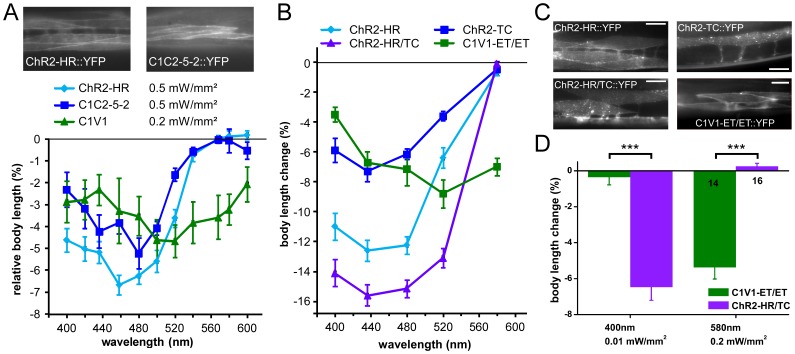
The C. elegans nervous system is particularly well suited for optogenetic analyses of circuit function: Essentially all connections have been mapped, and light can be directed at the neuron of interest in the freely moving, transparent animals, while behavior is observed. Thus, different nodes of a neuronal network can be probed for their role in controlling a particular behavior, using different optogenetic tools for photo-activation or -inhibition, which respond to different colors of light. As neurons may act in concert or in opposing ways to affect a behavior, one would further like to excite these neurons concomitantly, yet independent of each other. In addition to the blue-light activated Channelrhodopsin-2 (ChR2), spectrally red-shifted ChR variants have been explored recently. Here, we establish the green-light activated ChR chimera C1V1 (from Chlamydomonas and Volvox ChR1's) for use in C. elegans. We surveyed a number of red-shifted ChRs, and found that C1V1-ET/ET (E122T; E162T) works most reliable in C. elegans, with 540-580 nm excitation, which leaves ChR2 silent. However, as C1V1-ET/ET is very light sensitive, it still becomes activated when ChR2 is stimulated, even at 400 nm. Thus, we generated a highly efficient blue ChR2, the H134R; T159C double mutant (ChR2-HR/TC). Both proteins can be used in the same animal, in different neurons, to independently control each cell type with light, enabling a further level of complexity in circuit analyses.


Conflict of interest statement

Competing Interests: The authors have declared that no competing interests exist.

Figures

Figure 1
Figure 1. Survey of various ChR variants for red-shifted excitation in C. elegans muscle.
A) Upper panel, fluorescence micrographs. ChR2(H134R)::YFP (ChR2-HR), C1C2 5-2::YFP and C1V1::YFP (for expression pattern, see Fig. S5A), were expressed in body wall muscle cells and photostimulated (lower panel; light intensities and wavelength of band pass filters used are indicated). The resulting body length changes (contractions) were measured relative to the initial length of unstimulated animals. N = 7–24 animals were used for each experiment. B) ChR2-HR, ChR2-TC, ChR2-HR/TC and C1V1-ET/ET were expressed in muscles cells and the resulting body length changes in response to 0.2 mW/mm2 light at 5 different wavelengths were measured. N = 9–10 animals each. In A and B, absolute contractions (during light) were significantly different from the length before photostimulation for all data points, except for ChR2 and C1C2 at 560 and 568 nm. For a comparison of normalized data of the experiments in A and B, and analysis of statistically significant differences in contractions relative to those evoked by ChR2-HR, see Fig. S3. C) Expression patterns of the 4 proteins described in B, each fused to YFP, in body wall muscle cells. Scale bars are 20 µm. D) Light-evoked contractions of animals expressing C1V1-ET/ET or ChR2-HR/TC (individually) in body wall muscles were compared at 400 nm (0.01 mW/mm2) and 580 nm (0.2 mW/mm2). Each protein evokes strong contractions at its near-peak wavelength, but is not co-activated by the respective other wavelength. Number of animals is indicated. In A, B, D, mean body length changes and s.e.m. are displayed. In D, statistically significant differences after two-tailed Student’s t-test are indicated (***p<0.001).
Figure 2
Figure 2. C1V1-ET/ET and ChR2-HR evoke wavelength-dependent activity in mechanosensory neurons. A)
The six gentle touch mechanoreceptor neurons, their localization and morphology are shown. B) The effectiveness of C1V1-ET/ET and ChR2-HR, expressed in different strains, in evoking behavioral responses in mechanoreceptor neurons (backward escape, reversals) were compared, in response to 400 and 568 nm light of different intensities, as indicated. While C1V1-ET/ET can be specifically activated without co-activation of ChR2-HR at 568 nm, significant co-activation of C1V1-ET/ET at 400 nm is apparent. Displayed is the mean probability of backing in response to blue light stimuli (and s.e.m.), as described in Methods. Number of animals is indicated, and statistically significant differences were determined by two-tailed Student’s t-test, as indicated (***p<0.001).
Figure 3
Figure 3. (Co-)expression of ChR2-HR and C1V1-ET/ET in different cell types of the same animal.
A) ChR2-HR and C1V1-ET/ET were expressed in different cell types (GABAergic motor neurons or body wall muscle cells), either in different transgenic lines, or co-expressed in the same animal, as indicated in the pictograms. Fluorescence micrograph shows expression in the same animal of ChR2-HR::YFP in GABA neurons and C1V1-ET/ET::YFP in muscle cells. B) Body length changes (contraction or relaxation = elongation; given in relation to the initial body length of unstimulated animals) were measured in response to light of the indicated wavelengths and intensities. Residual contractions evoked by 400 nm light by C1V1-ET/ET in muscle could be counteracted, but not overcome, by ChR2-HR in GABA neurons. Displayed are mean body length changes and s.e.m. N = 10 animals of each strain were measured. Statistically significant differences were calculated by two-tailed Student’s t-test (n.s. = non significant; ***p<0.001).
Figure 4
Figure 4. ChR2-HR/TC in GABA neurons and C1V1-ET/ET in ACh neurons enable independent bi-modal light control of either cell type. A)
Innervation of muscle cells by GABAergic and cholinergic motor neurons, expressing ChR2-HR/TC and C1V1-ET/ET, respectively, in the same animal. B) Confocal fluorescence image of ChR2-HR/TC::YFP and C1V1-ET/ET::mCherry expressed in the ventral nerve cord motor neurons. C) Body length changes were measured in response to 400 or 580 nm light of the indicated intensities. Animals used expressed either ChR2-HR/TC in GABA neurons, or C1V1-ET/ET in cholinergic neurons, or both. When both proteins were expressed in the same animal, independent activation of both proteins and the respective neuron type can be achieved. Displayed are mean body length changes, relative to the initial length of the non-illuminated animals, and s.e.m. N = 12 animals of each strain were measured. Statistically significant differences were calculated by two-tailed Student’s t-test (n.s. = non significant; *p<0.05; **p<0.01; ***p<0.001).

References

    1. Fenno L, Yizhar O, Deisseroth K (2011) The development and application of optogenetics. Annu Rev Neurosci 34: 389–412. - PMC - PubMed
    1. Xu X, Kim SK (2011) The early bird catches the worm: new technologies for the Caenorhabditis elegans toolkit. Nat Rev Genet 12: 793–801. - PMC - PubMed
    1. Rein ML, Deussing JM (2012) The optogenetic (r)evolution. Mol Genet Genomics 287: 95–109. - PMC - PubMed
    1. Bernstein JG, Boyden ES (2011) Optogenetic tools for analyzing the neural circuits of behavior. Trends Cogn Sci 15: 592–600. - PMC - PubMed
    1. Nagel G, Szellas T, Kateriya S, Adeishvili N, Hegemann P, et al. (2005) Channelrhodopsins: directly light-gated cation channels. Biochem Soc Trans 33: 863–866. - PubMed

Publication types

MeSH terms