. 2012;7(10):e46903.
doi: 10.1371/journal.pone.0046903. Epub 2012 Oct 2.

Aphid gel saliva: sheath structure, protein composition and secretory dependence on stylet-tip milieu

Affiliations

Aphid gel saliva: sheath structure, protein composition and secretory dependence on stylet-tip milieu

Torsten Will et al. PLoS One. 2012.

Abstract

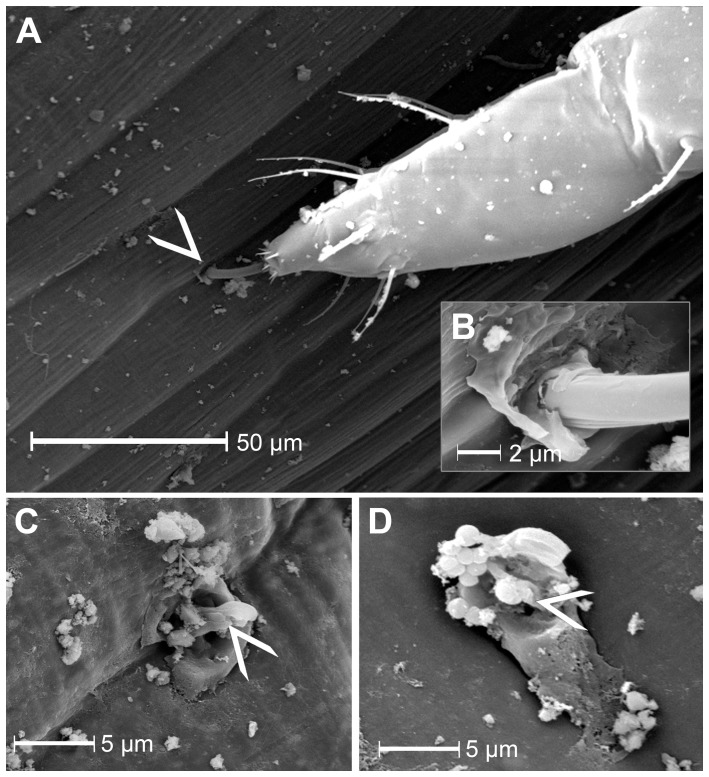
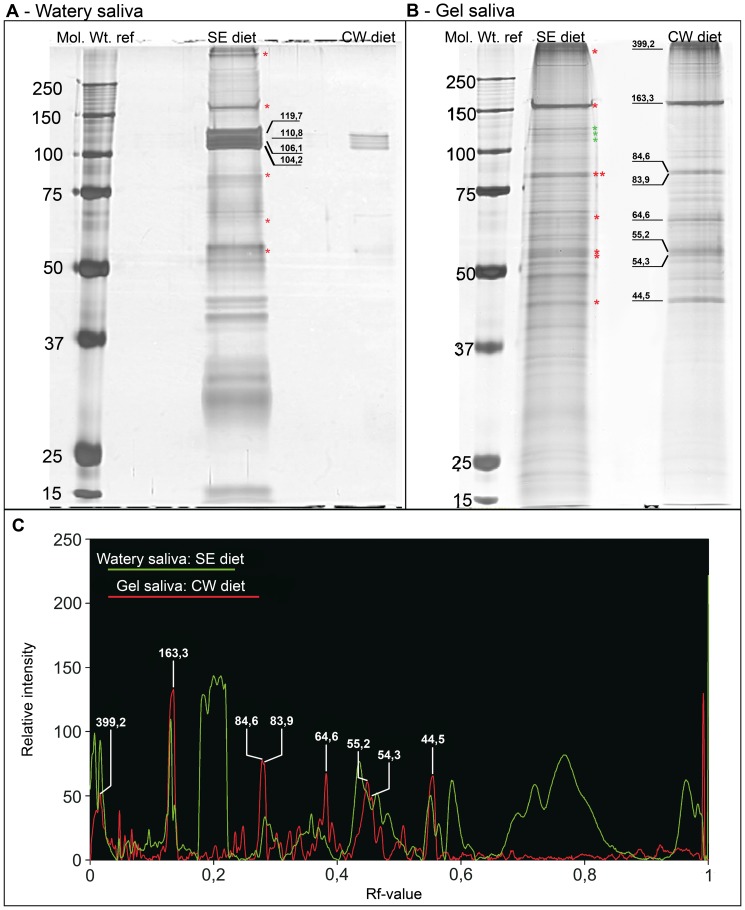
In order to separate and analyze saliva types secreted during stylet propagation and feeding, aphids were fed on artificial diets. Gel saliva was deposited as chains of droplets onto Parafilm membranes covering the diets into which watery saliva was secreted. Saliva compounds collected from the diet fluid were separated by SDS-PAGE, while non-soluble gel saliva deposits were processed in a novel manner prior to protein separation by SDS-PAGE. Soluble (watery saliva) and non-soluble (gel saliva) protein fractions were significantly different. To test the effect of the stylet milieu on saliva secretion, aphids were fed on various diets. Hardening of gel saliva is strongly oxygen-dependent, probably owing to formation of sulfide bridges by oxidation of sulphydryl groups. Surface texture of gel saliva deposits is less pronounced under low-oxygen conditions and disappears in dithiothreitol containing diet. Using diets mimicking sieve-element sap and cell-wall fluid respectively showed that the soluble protein fraction was almost exclusively secreted in sieve elements while non-soluble fraction was preferentially secreted at cell wall conditions. This indicates that aphids are able to adapt salivary secretion in dependence of the stylet milieu.


Conflict of interest statement

Competing Interests: The authors have declared that no competing interests exist.

Figures

Figure 1
Figure 1. Scanning electron microscopy of stylet penetration sites of M. euphorbiae.
A) The stylet entry point (arrowhead), which always seems to be located at the subsiding rims of anticlinal wall junctions between epidermal cells (N = 5), B) (magnified detail from A) is surrounded by a salivary flange. The saliva material partially covers the leaf surface around the stylet. C) After stylet retraction, the salivary flange appears to be plugged (N = 10). In the centre of the salivary sheath (arrowhead) saliva material shows a small elevation. Bacteria, spherical structures in the upper left region of the flange, reside on the gel saliva material. Growth of bacteria on the salivary flange material was observed regularly.
Figure 2
Figure 2. SEM micrographs of salivary sheaths from the aphid M. viciae formed under different oxidizing conditions.
Sheaths in each micrograph are entirely attached to the Parafilm membrane that covered the diet; the initial stylet penetration site in Fig. 2A, C and d is located in the lower left corner. A) A salivary sheath, formed under regular oxygen conditions, shows the typical pearl-necklace structure. The spherical parts of the gel saliva are formed by individual pulses of gel saliva and show clear delimitation from each other. The salivary sheath with a total length of approximately 52 µm tapers towards the tip and shows three short branches (arrowheads) in nearly regular intervals of 12.2; 16.9 and 13.1 µm (mean: 14.07 µm). The area within the white frame is zoomed (B) and shows a rough texture of the salivary sheath. A short branch is marked with an arrow head. C) A gel saliva sheath formed under oxygen-reduced conditions with less clear delimitation of the single saliva drops and a smoother texture than under oxygen conditions. The sheath structure appears to be more flat than under oxygen conditions. D) Addition of 1 mM DTT to low-oxygen ST-diet leads to a complete loss of the sheath structure. Potential remnants of the pearl-necklet appearance (shown exemplarily by white arrowheads) are visible at regular intervals inside the amorphous sheath.
Figure 3
Figure 3. Optical sectioning by CLSM through a salivary sheath of M. viciae.
The salivary sheath is attached along its whole length of 106 µm to the lower side of Parafilm membrane that covered a feeding chamber filled with ST diet. The site of stylet penetration through the Parafilm is located on the right end of the salivary sheath (right arrow). The sheath has a typical pearl-necklace like structure and narrows slightly towards the tip (left arrow). The empty stylet canal is visible inside the salivary sheath (indicated by arrowheads) with exception of the very first segment (right arrow).
Figure 4
Figure 4. Separation and comparison of saliva protein fractions from M. viciae collected in SE – and CW-diet.
Soluble and non-soluble protein fraction of saliva concentrate collected from SE-diet and CW-diet were separated in a 10% separation gel. Gels were silver stained. Lane 1 of each gel contains marker proteins Molecular weights (kDa) are given on the left. A) The soluble fraction from SE-diet shows 45 protein bands while in samples from CW-diet only 6 protein bands are detected. All latter protein bands match bands from the SE-diet but have a much lower abundance. B) Non-soluble fraction samples have fewer protein bands in CW-diet (lane 3, n = 66) than in SE-diet (lane 2, n = 87). Matching bands of lanes 2 and 3 show comparable intensities. The most intense protein bands (MWs are given in B) of the non-soluble fraction from CW-diet are used as reference proteins for this saliva fraction and are labeled with their MWs. Protein bands with corresponding MW in other lanes are marked with red asterisks. Protein bands of insoluble fraction that correspond to soluble fraction from SE-diet (A) are labeled with green asterisks and MWs are given in A. C) To discriminate protein bands of non-soluble fraction (red graph) from those of soluble fraction (green graph), soluble fraction from SE-diet (A, lane 2) was compared with non-soluble fraction from CW-diet (B, lane 3). The relative pixel intensity value (arbitrary units) is plotted against the retardation factor (Rf-value). Most intense proteins from non-soluble fraction (MWs are given), all of which have a relative pixel intensity >50, show a coincidence with 6 protein bands in the soluble fraction collected from SE-diet (red asterisks). Specific proteins from soluble fraction with a relative intensity of more than 50 (RF-value of approx. 0.2 (MWs in A and green asterisks in B) and higher 0.57), are only slightly visible in non-soluble fraction from CW-diet (C).

References

    1. Hewer A, Will T, van Bel AJE (2010) Plant cues for aphid navigation in vascular tissues. J Exp Biol 213: 4030–4042. - PubMed
    1. Hewer A, Becker A, van Bel AJE (2011) An aphid's Odyssey. The cortical quest for the vascular bundle. J Exp Biol 214: 3868–3879. - PubMed
    1. Martin B, Collar JL, Tjallingii WF, Fereres A (1997) Intracellular ingestion and salivation by aphids may cause the acquisition and inoculation of non-persistently transmitted plant viruses. J Gen Virol 78: 2701–2705. - PubMed
    1. Powell G (2005) Intracellular salivation is the aphid activity associated with inoculation of non-persistently transmitted viruses. J Gen Virol 86: 469–472. - PubMed
    1. Ponsen MB (1972) The site of potato leafroll virus multiplication in its vector, Myzus persicae . Mededelingen van de Landbouwhogeschool Wageningen 16: 1–147.

MeSH terms