. 2012;7(10):e47109.
doi: 10.1371/journal.pone.0047109. Epub 2012 Oct 8.

Novel agents targeting the IGF-1R/PI3K pathway impair cell proliferation and survival in subsets of medulloblastoma and neuroblastoma

Affiliations

Novel agents targeting the IGF-1R/PI3K pathway impair cell proliferation and survival in subsets of medulloblastoma and neuroblastoma

Anna Wojtalla et al. PLoS One. 2012.

Abstract

The receptor tyrosine kinase (RTK)/phosphoinositide 3-kinase (PI3K) pathway is fundamental for cancer cell proliferation and is known to be frequently altered and activated in neoplasia, including embryonal tumors. Based on the high frequency of alterations, targeting components of the PI3K signaling pathway is considered to be a promising therapeutic approach for cancer treatment. Here, we have investigated the potential of targeting the axis of the insulin-like growth factor-1 receptor (IGF-1R) and PI3K signaling in two common cancers of childhood: neuroblastoma, the most common extracranial tumor in children and medulloblastoma, the most frequent malignant childhood brain tumor. By treating neuroblastoma and medulloblastoma cells with R1507, a specific humanized monoclonal antibody against the IGF-1R, we could observe cell line-specific responses and in some cases a strong decrease in cell proliferation. In contrast, targeting the PI3K p110α with the specific inhibitor PIK75 resulted in broad anti-proliferative effects in a panel of neuro- and medulloblastoma cell lines. Additionally, sensitization to commonly used chemotherapeutic agents occurred in neuroblastoma cells upon treatment with R1507 or PIK75. Furthermore, by studying the expression and phosphorylation state of IGF-1R/PI3K downstream signaling targets we found down-regulated signaling pathway activation. In addition, apoptosis occurred in embryonal tumor cells after treatment with PIK75 or R1507. Together, our studies demonstrate the potential of targeting the IGF-1R/PI3K signaling axis in embryonal tumors. Hopefully, this knowledge will contribute to the development of urgently required new targeted therapies for embryonal tumors.


Conflict of interest statement

Competing Interests: The authors have declared that no competing interests exist.

Figures

Figure 1
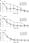
Figure 1. The effect of R1507 on cell proliferation of NB and MB cells.
A panel of NB cell lines (A) and MB cell lines (B) were incubated with increasing concentrations of the antibody R1507 inhibiting the IGF-1R in serum-containing medium. Cell viability was assessed using the MTS assay after 2 days. The data represent the mean with SD from at least 6 replicates and 3 independent experiments.
Figure 2
Figure 2. Sensitivity to R1507 and PIK75, and presence of IGFR in neuroblastoma cell lines LAN1 and LAN1R, a LAN1 cell line resistant to doxorubicin.
(A) R1507 treatment for 48 hours. (B) PIK75 treatment for 48 hours. Error bars represent ±S.D. of means from 3 experiments, each with 3 replicates, except that there was only one experiment with 500 nM PIK75 in B. (C) Western blot analysis of components of the IGF-1R/PI3K pathway in LAN1 and LAN1R whole cell extracts. Src was used as internal loading control. (D) Transfection of LAN1 cells with siRNA targeting the IGF-1R (non-targeting siRNA was used as control. Expression levels of the IGF-1R were assessed by Western blot analysis in LAN1 whole cell lysates after 96 h). Cell proliferation in LAN1 cells upon IGF-1R silencing was assessed in absence or presence of R1507 after 96h by MTS. (*p<0.05).
Figure 3
Figure 3. Cell proliferation of NB and MB cells after inhibition of the PI3K p110α.
A panel of NB cell lines (A) and MB cell lines (B) were incubated with increasing concentrations of the specific pharmacological PI3K p110α inhibitor PIK75 in serum-containing medium. Cell viability was assessed using the MTS assay after 2 (NB) or 3 (MB) days. The data represent the mean with SD from at least four replicates and 1–3 independent experiment.
Figure 4
Figure 4. PI3K and IGF-1R inhibition impair receptor activation and downstream signaling.
The NB cells SH-SY5Y (A), LAN1 (B); WAC2 (C) and the MB cells PFSK (D) grown in serum-containing medium were incubated with increasing concentrations of the PI3K p110α inhibitor PIK75 and the IGF-1R antibody R1507. After 24 hours the cells were harvested and whole cell lysates analysed by SDS-PAGE and Western blotting for the proteins indicated. Serum-starved DAOY (E) or LAN1 (F) cells were pre-treated with vehicle or R1507 at the concentrations indicated for 1h and stimulated with IGF-1 (50 ng/ml or 100 ng/ml) for 10 min at 37°C. Cell lysates were analysed by SDS-PAGE and Western Blot for phosphorylated IGF-1R beta and total receptor.
Figure 5
Figure 5. Treatment of NB cells with R1507 or PIK75 in presence of chemotherapy results in additive effects.
NB cells grown in serum-containing medium were incubated with the IGF-1R antibody R1507 (0.1 μg/ml) (A) or the PI3K inhibitor PIK75 (0.05 μM) (B+C) in presence or absence of cisplatin (1 μM), etoposide (1 μM), or doxorubicin (0.1 μM). Cell proliferation was assessed using the MTS assay after 48 h. The data represent the mean of 8 replicates with SD from 3 independent experiments. (*p<0.05).
Figure 6
Figure 6. Additive/sensitization effects after combinatorial treatment with R1507 and chemotherapy in MB cells.
The MB cell lines PFSK (A), UW228 (B) and DAOY (C) grown in serum-containing medium were incubated with increasing concentrations of cisplatin in presence or absence of the IGF-1R antibody R1507 (25 µg/ml or 50 µg/ml). Cell proliferation was assessed using the MTS assay after 48 h. The data represent the mean of 6 replicates with SD from 3 independent experiments.
Figure 7
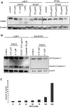
Figure 7. Apoptosis upon PI3K inhibition.
(A+B) The NB cell lines LAN1 (A, left panel; B, left panel) and SH-SY5Y (B, right panel) as well as the MB cell line PFSK (A, right panel) grown in serum-containing medium were incubated with increasing concentrations of the PI3K p110α inhibitor PIK75 and the IGF-1R antibody R1507, or cisplatin. After 24 hours the cells were harvested and whole cell lysates analysed by SDS-PAGE and Western blotting for the proteins indicated. (C) The NB cell line WAC2 grown in serum-containing medium was incubated with increasing concentrations of the PI3K p110α inhibitor PIK75. Caspase 3/7 activity was assessed using the Caspase 3/7 Glo assay after 48 h.
Figure 8
Figure 8. Targeted therapies in medulloblastoma.
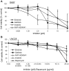
The MB cell lines Daoy (A) and UW228 (B) were incubated with increasing concentrations of the EGFR inhibitors gefinitib or erlotinib, the Abl inhibitor imatinib, the IGF-1R inhibitor NVP-AEW541 and the mTOR inhibitor rapamycin. Cell proliferation was assessed using the MTS assay after 72 h. The data represent the mean of 6 replicates with SD from 3 independent experiments.
Figure 9
Figure 9. Effects of R1507 in combination with targeted therapies in medulloblastoma.
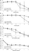
The R1507-insensitive cell line DAOY was incubated with increasing concentrations of the EGFR inhibitor gefinitib (A), the Abl inhibitor imatinib (B), the IGF-1R inhibitor NVP-AEW541 (C) and the mTOR inhibitor rapamycin (D) in presence or absence of the IGF-1R antibody R1507. Cell proliferation was assessed using the MTS assay after 72 h. The data represent the mean of 6 replicates with SD from 3 independent experiments.

References

    1. Schwab M, Praml C, Amler LC (1996) Genomic instability in 1p and human malignancies. Genes Chromosomes Cancer 16: 211–229. - PubMed
    1. Schwab M, Alitalo K, Klempnauer KH, Varmus HE, Bishop JM, et al. (1983) Amplified DNA with limited homology to myc cellular oncogene is shared by human neuroblastoma cell lines and a neuroblastoma tumour. Nature 305: 245–248. - PubMed
    1. Plantaz D, Vandesompele J, Van Roy N, Lastowska M, Bown N, et al. (2001) Comparative genomic hybridization (CGH) analysis of stage 4 neuroblastoma reveals high frequency of 11q deletion in tumors lacking MYCN amplification. Int J Cancer 91: 680–686. - PubMed
    1. Van Roy N, Van Limbergen H, Vandesompele J, Van Gele M, Poppe B, et al. (2001) Combined M-FISH and CGH analysis allows comprehensive description of genetic alterations in neuroblastoma cell lines. Genes Chromosomes Cancer 32: 126–135. - PubMed
    1. Van Roy N, Jauch A, Van Gele M, Laureys G, Versteeg R, et al. (1997) Comparative genomic hybridization analysis of human neuroblastomas: detection of distal 1p deletions and further molecular genetic characterization of neuroblastoma cell lines. Cancer Genet Cytogenet 97: 135–142. - PubMed

Publication types

MeSH terms

Substances