. 2012;7(10):e47142.
doi: 10.1371/journal.pone.0047142. Epub 2012 Oct 8.

Cthrc1, a novel circulating hormone regulating metabolism

Affiliations

Cthrc1, a novel circulating hormone regulating metabolism

J Patrizia Stohn et al. PLoS One. 2012.

Abstract

Background: We discovered the gene Collagen Triple Helix Repeat Containing 1 (Cthrc1) and reported its developmental expression and induction in adventitial cells of injured arteries and dermal cells of skin wounds. The role of Cthrc1 in normal adult tissues has not yet been determined.

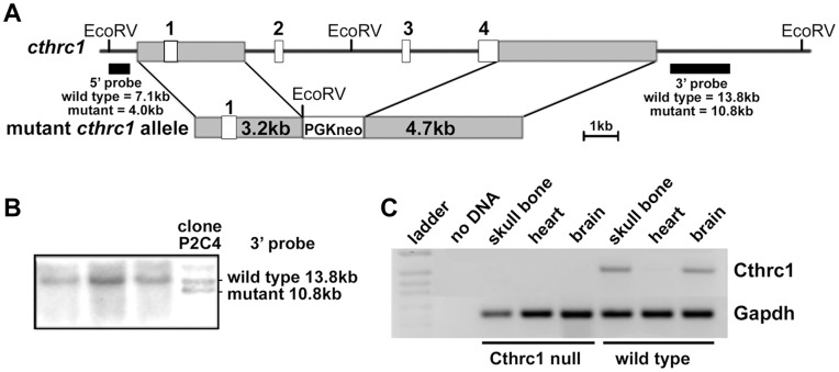
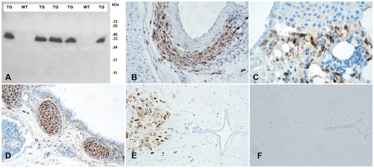
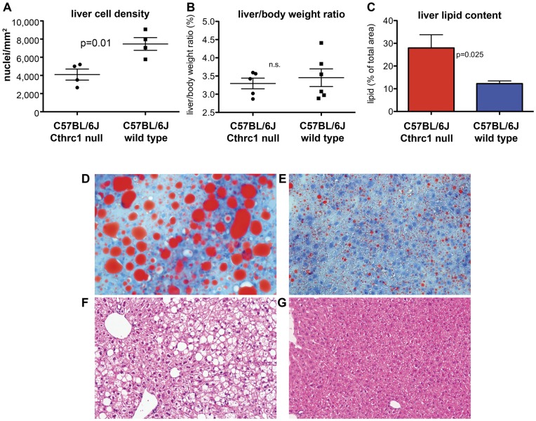
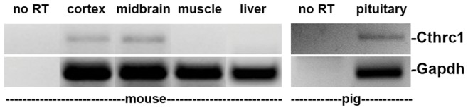
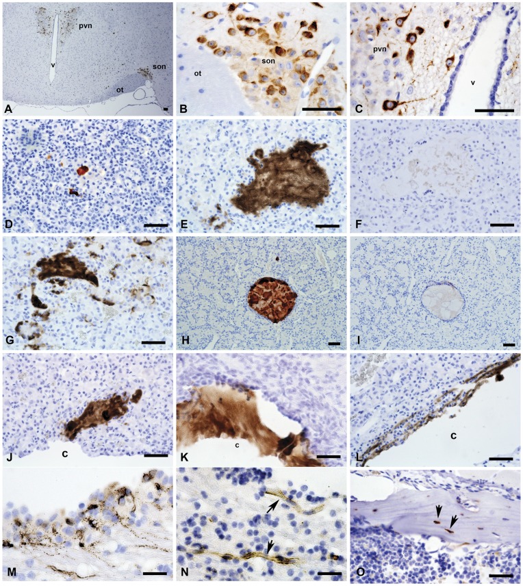
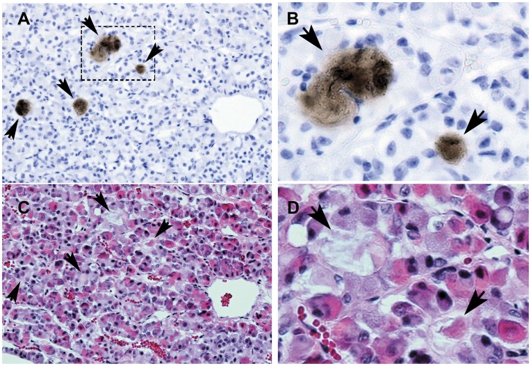
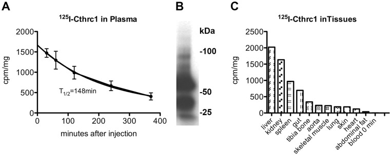
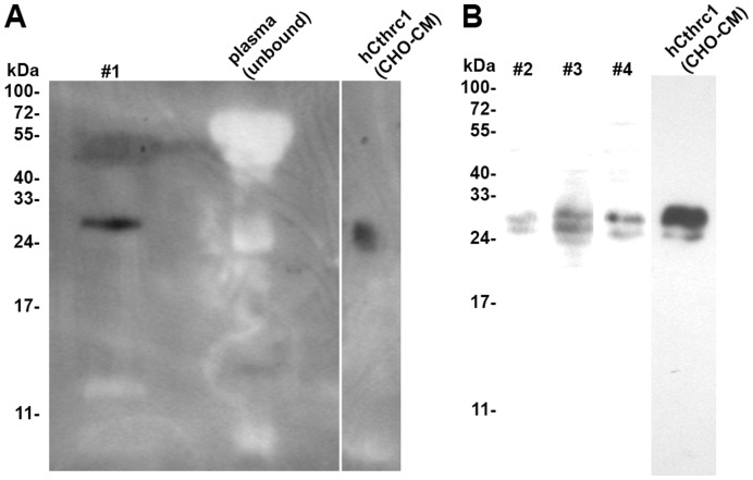
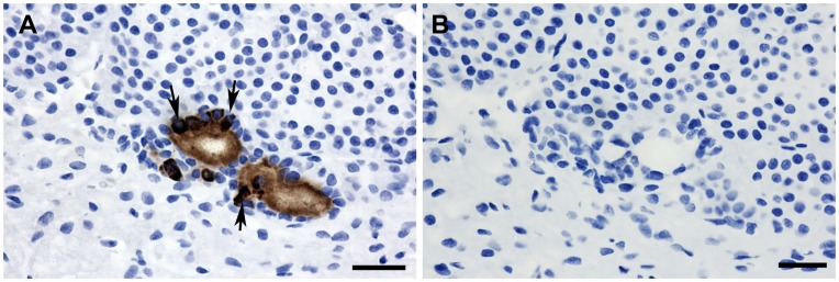
Methodology/principal findings: We generated mutant mice with a novel Cthrc1 null allele by homologues recombination. Cthrc1 null mice appeared developmentally normal. On the C57BL/6J background, livers from Cthrc1 null mice accumulated vast quantities of lipid, leading to extensive macrovesicular steatosis. Glycogen levels in skeletal muscle and liver of Cthrc1 null mice on the 129S6/SvEv background were significantly increased. However, Cthrc1 expression is not detectable in these tissues in wild-type mice, suggesting that the lipid and glycogen storage phenotype may be a secondary effect due to loss of Cthrc1 production at a distant site. To investigate potential hormonal functions of Cthrc1, tissues from adult mice and pigs were examined for Cthrc1 expression by immunohistochemistry with monoclonal anti-Cthrc1 antibodies. In pigs, Cthrc1 was detected around chromophobe cells of the anterior pituitary, and storage of Cthrc1 was observed in colloid-filled follicles and the pituitary cleft. Pituitary follicles have been observed in numerous vertebrates including humans but none of the known pituitary hormones have hitherto been detected in them. In C57BL/6J mice, however, Cthrc1 was predominantly expressed in the paraventricular and supraoptic nucleus of the hypothalamus but not in the posterior pituitary. In human plasma, we detected Cthrc1 in pg/ml quantities and studies with (125)I-labeled Cthrc1 revealed a half-life of 2.5 hours in circulation. The highest level of Cthrc1 binding was observed in the liver.

Conclusions: Cthrc1 has characteristics of a circulating hormone generated from the anterior pituitary, hypothalamus and bone. Hormonal functions of Cthrc1 include regulation of lipid storage and cellular glycogen levels with potentially broad implications for cell metabolism and physiology.


Conflict of interest statement

Competing Interests: The authors have declared that no competing interests exist.

Figures

Figure 1
Figure 1. Generation of Cthrc1 null mice.
(A) Cthrc1 gene targeting schematic showing replacement of exons 2–4 with a PGK-neo cassette. (B) Embryonic stem cell targeting with Southern blot analysis of genomic DNA from ES cell clones shows a targeted clone. (C) RT-PCR shows lack of Cthrc1 transcripts in RNA preparations of skull bone and whole brain of adult Cthrc1 mutants.
Figure 2
Figure 2. Characterization of rabbit monoclonal anti-Cthrc1 (Vli-55) antibody.
(A) Western blot analysis of plasma from Cthrc1 transgenic mice (TG) and their negative wild type littermates (WT). (B) Cthrc1 immunohistochemistry shows expression in adventitial cells of the remodeling renal artery following angiotensin II infusion, (C) dermal cells in 7 day old skin wounds, and (D) embryonic cartilage (E17.5). (E) Cthrc1 expression is restricted to the midbrain in wild type 129S6/SvEv mice and (F) no expression is detected in the midbrain of a corresponding Cthrc1 null mouse.
Figure 3
Figure 3. Normal growth but increased glycogen storage in liver and skeletal muscle of 129S6/SvEv Cthrc1 null mice.
(A) Differences in litter size were not significantly different (n.s.). (B) Growth curves for females and (C) males were similar (n = 5–33 mice per time point). (D, E) Total body fat expressed as a percentage of body weight was similar in 8 week old female mice among mutants and wild type mice. (F) Blood triglyceride levels in 3–4 month old male and female mice were similar. (G) Glycogen storage in skeletal muscle and (H) liver is significantly increased in Cthrc1 null mice on the 129S6/SvEv background (males and females, 3–5 months of age). (I) Glucose handling is normal in Cthrc1 null mice (n = 8 per group, mixed gender, 3–4 months old). (J) Reduced insulin sensitivity was observed in Cthrc1 null mice (n = 8 per group, mixed gender, 3–4 months old).
Figure 4
Figure 4. Fatty liver phenotype in C57BL/6J Cthrc1 null mice.
(A) Cell density in livers of wild type and Cthrc1 null mice (n = 4 per group). (B) Liver to body weight ratio was similar among groups. (C) Lipid content in liver was determined by image analysis on oil red O stained sectins of 7 month old mixed gender Cthrc1 null and wild type mice (n = 6 per group). (D) Oil red O stain of a liver section from a Cthrc1 null mouse shows macrovesicular and microvesicular steatosis and (E) only minor lipid accumulations in a wild type mouse. (F) Hematoxylin & eosin stain of a liver section from a Cthrc1 null mouse and (G) a wild type mouse.
Figure 5
Figure 5. Cthrc1 expression in the pig pituitary but absence in adult mouse liver and skeletal muscle.
RT-PCR with Cthrc1 specific primer pair was performed on the indicated mouse and pig RNA samples.
Figure 6
Figure 6. Cthrc1 immunohistochemistry of the brain and pituitary gland from mouse and pig.
Sections of mouse (A–D) and pig (E–N) brain and pituitary were immunostained for Cthrc1 expression: (A) Low power view of a coronal section of the hypothalamus from a C57BL/6J mouse showing Cthrc1 immunoreactive cells in the paraventricular nucleus (pvn) and the supraoptic nucleus (son), ot = optic tract, v = third ventricle. (B) High power view of supra optic nucleus and (C) paraventricular nucleus. (D) Cthrc1 positive follicle in the anterior lobe of a 7 month old C57BL6/J male, (E) extensive accumulations of Cthrc1 in the anterior lobe of a pig pituitary, and (F) pre-absorption of antibody completely eliminates staining on an adjacent section. (G) Cytoplasmic localization of Cthrc1 in cells of the anterior lobe indicates expression. (H–I) are serial sections of a typical colloid-filled follicle of the anterior pituitary with (H) showing extensive immunoreactivity, (I) which is completely eliminated by pre-absorbing the antibody with peptide antigen (H). Also note the encapsulation of the follicle by folliculostellate cells. (J, K) Cthrc1 localization in the pituitary cleft (c) and (L) canaliculi connecting to the cleft. (M) An isolated area of cells in the paraventricular zone of the lateral ventricle expresses Cthrc1 (note granular appearance), and (N) nearby small vessels (arrows) contain Cthrc1. (O) Cthrc1 is expressed by some osteocytes (arrows) and osteoblasts in adult mouse bone. Scale bar = 50 µm.
Figure 7
Figure 7. Cthrc1 accumulation in areas surrounded by chromophobe cells of the anterior pig pituitary.
Adjacent sections were stained for Cthrc1 (A) and morphology (C, H&E). (B) and (D) are enlarged images of the boxed area shown in (A). Arrows indicate areas of Cthrc1 accumulation. Scale bar = 50 µm.
Figure 8
Figure 8. Pharmacokinetic properties of Cthrc1.
(A) Clearance curve of 125I-Cthrc1 injected intra-arterially into Cthrc1 null mice. The half-life of the protein was calculated to be 146 min. (B) SDS-PAGE and autoradiographic analysis of plasma 30 min after 125I-Cthrc1 injection. Monomeric, dimeric and higher molecular weight forms of Cthrc1 were detectable. (C) 125I-Cthrc1 content in various tissues 6 h after injection.
Figure 9
Figure 9. Cthrc1 detection in human plasma.
A pull-down assay with monoclonal Cthrc1 antibody coupled to magnetic beads was performed on human plasma samples (#1–#4). (A) The eluate was analyzed by Western blotting with HRP-conjugated anti-Cthrc1 antibody, conditioned medium from hCthrc1 expressing CHO-K1 cells was used as a positive control. No Cthrc1 was detectable in the unbound plasma fraction (note absence of non-specific bands). (B) The eluates from plasma samples #2–#4 were immunoblotted with biotin-conjugated anti-Cthrc1 antibody followed by HRP-conjugated streptavidin.
Figure 10
Figure 10. Cthrc1 secretion is cell type dependent.
Detection of Cthrc1 in conditioned medium (CM) and cell lysate (CL) of CHO-K1 or HEK293T cells 72 h after transfection with a Cthrc1 expression vector. Note the absence of Cthrc1 in the CM of HEK293T cells.
Figure 11
Figure 11. Isolated cells in the rat pituitary express Cthrc1.
(A) Cthrc1 immunohistochemistry on pituitary glands from three month old male Sprague Dawley rats identified Cthrc1 expression by isolated cells. Cytoplasmic immunoreactivity is clearly detectable in cells adjacent to extracellular accumulations of Cthrc1 (arrows), suggesting Cthrc1 synthesis by these cells. (B) Pre-absorption of antibody with peptide antigen completely eliminates staining on an adjacent section. Scale bar = 50 µm.

References

    1. Pyagay P, Heroult M, Wang Q, Lehnert W, Belden J, et al. (2005) Collagen triple helix repeat containing 1, a novel secreted protein in injured and diseased arteries, inhibits collagen expression and promotes cell migration. Circ Res 96: 261–268. - PubMed
    1. Yamamoto S, Nishimura O, Misaki K, Nishita M, Minami Y, et al. (2008) Cthrc1 selectively activates the planar cell polarity pathway of Wnt signaling by stabilizing the Wnt-receptor complex. Dev Cell 15: 23–36. - PubMed
    1. Kimura H, Kwan KM, Zhang Z, Deng JM, Darnay BG, et al. (2008) Cthrc1 is a positive regulator of osteoblastic bone formation. PLoS ONE 3: e3174. - PMC - PubMed
    1. LeClair RJ, Durmus T, Wang Q, Pyagay P, Terzic A, et al. (2007) Cthrc1 is a novel inhibitor of transforming growth factor-beta signaling and neointimal lesion formation. Circ Res 100: 826–833. - PubMed
    1. Ogawa S, Couch EF, Kubo M, Sakai T, Inoue K (1996) Histochemical study of follicles in the senescent porcine pituitary gland. Arch Histol Cytol 59: 467–478. - PubMed

Publication types

LinkOut - more resources