. 2012;7(10):e47187.
doi: 10.1371/journal.pone.0047187. Epub 2012 Oct 9.

Cultivation and characterization of cornea limbal epithelial stem cells on lens capsule in animal material-free medium

Affiliations

Cultivation and characterization of cornea limbal epithelial stem cells on lens capsule in animal material-free medium

Réka Albert et al. PLoS One. 2012.

Abstract

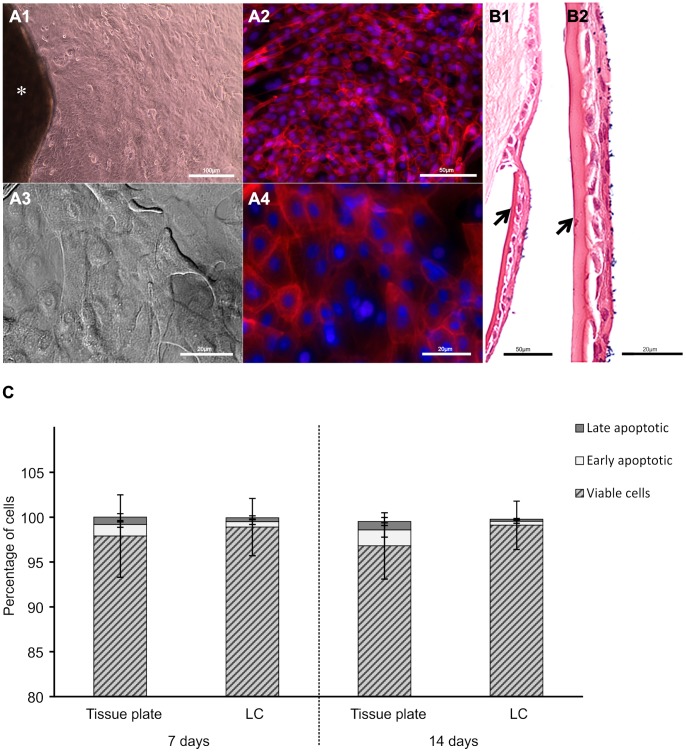
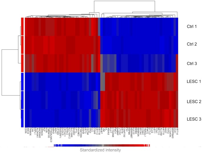
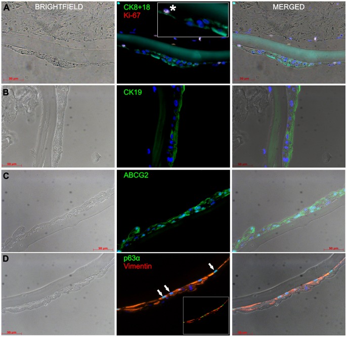
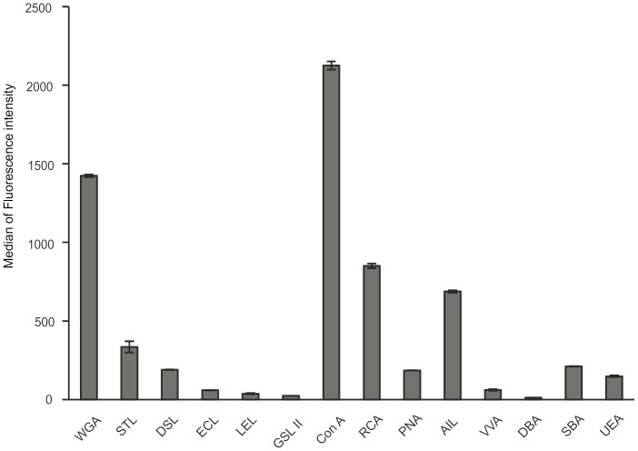
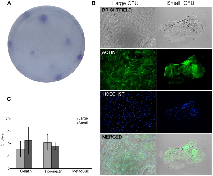
A simple, reproducible, animal-material free method for cultivating and characterizing cornea limbal epithelial stem cells (LESCs) on human lens capsule (LC) was developed for future clinical transplantation. The limbal tissue explants (2 × 2 × 0.25 mm) were harvested from 77 cadavers and expanded ex vivo on either cell culture plates or LC in medium containing human serum as the only growth supplement. Cell outgrowth at the edge of the explants was observed within 24 hours of cultivation and achieved viable outgrowth (>97% viability as measured by MTT assay and flow cytometry) within two weeks. The outgrowing cells were examined by genome-wide microarray including markers of stemness (p63α, ABCG2, CK19, Vimentin and Integrin α9), proliferation (Ki-67), limbal epithelial cells (CK 8/18 and 14) and differentiated cornea epithelial cells (CK 3 and 12). Immunostaining revealed the non-hematopoietic, -endothelial and -mesenchymal stem cell phenotype of the LESCs and the localization of specific markers in situ. Cell adhesion molecules, integrins and lectin-based surface carbohydrate profiling showed a specific pattern on these cells, while colony-formation assay confirmed their clonal potency. The LESCs expressed a specific surface marker fingerprint (CD117/c-kit, CXCR4, CD144/VE-Cadherin, CD146/MCAM, CD166/ALCAM, and surface carbohydrates: WGA, ConA, RCA, PNA and AIL) which can be used for better localization of the limbal stem cell niche. In summary, we report a novel method combining the use of a medium with human serum as the only growth supplement with LC for cultivating, characterizing and expanding cornea LESCs from cadavers or alternatively from autologous donors for possible treatment of LESC deficiency.


Conflict of interest statement

Competing Interests: The authors have declared that no competing interests exist.

Figures

Figure 1
Figure 1. Cultivation and viability of LESCs.
Limbal graft (*) cultured on cell culture plate (A) or human LC (B) showing outgrowth of cells with epithelial morphology within 24 hrs of cultivation (image shown represents a 3 day cell outgrowth, A1 and A3 are bright field images, A2 and A4 are immunofluorescent images of actin cytoskeleton (red) and nucleus (blue)). Hematoxylin & Eosin staining of LESCs grown on LC (arrows) forming stratified epithelial layer at day 7 (B1 and B2). Cell viability and death of the cultured LESCs (viable cells (striped bar), early apoptotic or annexin V-FITC+ cells (light gray bar); late apoptotic or annexin V-FITC/propidium iodide+ cells (dark gray bar)) (C). Data shown are mean ± S.D (n = 3, Scale bars: 100 µm A1, 50 µm A2, 20 µm A3–4; 50 µm B1, 20 µm B2).
Figure 2
Figure 2. Transcriptional profiling in LESCs.
Heatmap of the transcripts and functional clustering of 67 genes selected on the basis of their high or low FC or previously documented relation to LESCs (n = 3, p<0.01). Red and blue colors indicate high and low expression, respectively.
Figure 3
Figure 3. Expression of epithelial-, stemness- and proliferation specific markers in LESCs grown on human LC measured by immunoflourescence staining.
Immunohistochemistry was performed to detect the (co)-expression of CK8/18/Ki-67, CK19, ABCG2, Vim/p63α in the LESCs grown on human LC (Left column: bright field-; Center: immunofluorescent; Right column: merged image; Colors on the text correspond to the color of the marker examined, while all nuclei are stained blue with DAPI; Insert: shows co-localization of CK8+18 and Ki-67 and (*) refers to the different staining pattern in the region shown; Arrows: show expression of Vimentin in the basal cells; the images are representative of at least 3 independent experiments, scale bar: 50 µm).
Figure 4
Figure 4. Expression of carbohydrate molecules on the surface of LESCs.
Lectins-based staining of carbohydrate specific molecules on the surface of LESCs. For abbreviations used see Table 2. (Data shown are mean ± S.D. of the median of fluorescence intensity, n = 3).
Figure 5
Figure 5. Colony-forming potential of LESCs.
The LESCs were cultured at clonal density of 3000 cells/cm2 and early epithelial holoclone-like colony formation was recorded at day 7 of culture. LESCs formed colony forming units on Gelatin and Fibronectin coated plates as stained by crystal violet (0.5% w/v) (A). The colony forming unit (CFU)-forming cells were stained for actin (phalloidin-FITC, green) and Hoechst 33342 (blue, nuclear). The CFUs could be divided into two groups: large CFUs containing >50 cells, and small CFUs containing <50 LSCs. No significant difference in the CFU types on Gelatin and Fibronectin matrices was found, while MethoCult matrix-grown LESC formed no colonies at all (B). (Data shown are mean ± S.D., n = 3).

References

    1. Davanger M, Evensen A (1971) Role of the pericorneal papillary structure in renewal of corneal epithelium. Nature 229: 560–561. - PubMed
    1. Schermer A, Galvin S, Sun TT (1986) Differentiation-related expression of a major 64K corneal keratin in vivo and in culture suggests limbal location of corneal epithelial stem cells. J Cell Biol 103: 49–62. - PMC - PubMed
    1. Tseng SC (1989) Concept and application of limbal stem cells. Eye (Lond) 3 (Pt 2): 141–157. - PubMed
    1. Schlötzer-Schrehardt U, Kruse FE (2005) Identification and characterization of limbal stem cells. Exp Eye Res 81: 247–264. - PubMed
    1. Notara M, Alatza A, Gilfillan J, Harris AR, Levis HJ, et al. (2010) In sickness and in health: Corneal epithelial stem cell biology, pathology and therapy. Exp Eye Res 90: 188–195. - PubMed

Publication types