. 2009 Jan;4(1):40-58.

Limbal stem cells in review

Affiliations

Limbal stem cells in review

Marzieh Ebrahimi et al. J Ophthalmic Vis Res. 2009 Jan.

Abstract

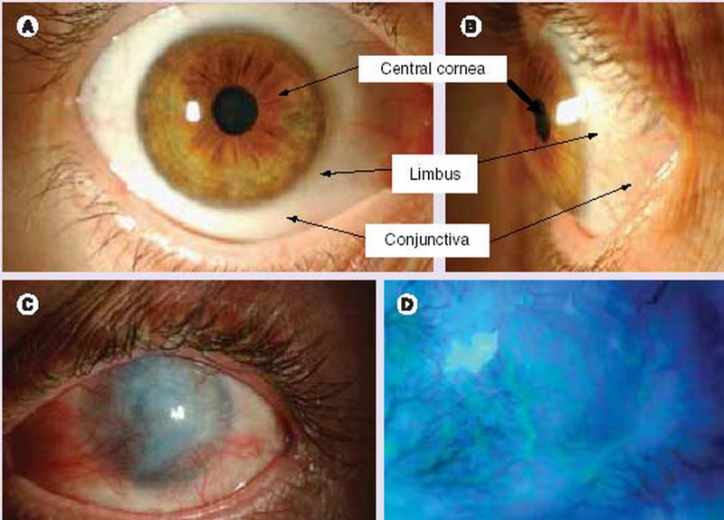
The ocular surface consists of two distinct types of epithelial cells; conjunctival and corneal. Although anatomically continuous, these epithelia comprise two distinct cell populations. Corneal stem cells are located at the limbus. The microenvironment of the limbus is important in maintaining "stemness" of the stem cells and also acts as a barrier to conjunctival epithelial cells preventing them from migration onto the corneal surface.Damage to the limbus results in varying degrees of limbal stem cell deficiency with characteristic clinical features including conjunctivalization of the cornea. Regenerative management of corneal conjunctivalization utilizing stem cells comprises of two approaches; limbal auto- or allografts by using existing stem cells and induction and regeneration of ocular tissues from embryonic stem cells. Herein, we review stem cells and limbal stem cells in particular, types of epithelial cells in the cornea, markers of corneal epithelial cells in different stages, as well as the current approach to corneal epithelial regeneration.

Keywords: Corneal; Epithelium; Limbus Corneae; Stem Cells.


Figures

Figure 1
Figure 1
The limbal palisades of Vogt.Palisades of Vogt (arrow) are easily recognized in the human limbus (A). Such a unique pigmented structure can be identified on the flat mount preparation of dispase-isolated human limbal epithelial sheets (B). In dark-skin donors, palisades of Vogt are pigmented (C, arrow). Under high magnification, these areas of limbus appear undulated (D, stars). Hematoxylin staining highlights higher stratification and further undulation of the limbal epithelium, and the underlying limbal stroma demonstrates high cellularity and vascularity (E, arrow shows blood vessel). The bar represents 500 μm in A and B, 200 μm in C and 50 μm in D.
Figure 2
Figure 2
Hypothetical diagram of the limbal stem cell niche. Limbal epithelial stem cells are located at the basement membrane of the limbus. In this epithelial level,there are several other cell types in the vicinity such as the immediate progeny, i.e., early transient amplifying cells (eTACs), melanocytes, and Langerhans’ cells. It remains to be determined whether these cell types act as niche cells. It is believed that eTACs are destined for progeny production by differentiating into late TACs located at the basement membrane of the cornea which consecutively differentiate into suprabasal post-mitotic cells, and finally into superficial terminally differentiated cells. The limbal basement membrane separates the epithelium from the underlying stroma. Limbal stroma contains mesenchymal cells, which may also serve as niche cells. Limbal stroma is highly innervated and vascularized; however the role of nerves and blood vessels in the niche remains to be defined.
Figure 3
Figure 3
Types of cell junctions (A), electron-microscopic location of cell junctions in cells (B),– and immunostaining of connexin-43 in limbal stem cell culture (C).
Figure 4
Figure 4
Immunofluorescent staining for connexin 43 in the cornea and limbus (arrows) (×100).
Figure 5
Figure 5
Expression of p63 in the epithelium of rabbit limbus (A) and cornea (B).
Figure 6
Figure 6
Immunostaining for cytokeratine 3 (K3) in the center of the cornea (Ebrahimi et al 2006; unpublished).K3 is expressed in the suprabasal (green) but not in the basal layer of the limbus or cornea.
Figure 7
Figure 7
Immunofluorescent staining for E-cadherin and P-cadherin expression on frozen sections of the human limbus. The arrows point to positively or negatively labeled basal cells at the limbus (×100).
Figure 8
Figure 8
Immunofluorescent staining for integrins expression on frozen sections of the human limbus. The arrows point to positively or negatively labeled basal cells at the limbus (×100).
Figure 9
Figure 9
Immunofluorescent staining for Growth factor receptors on frozen sections of the human limbus. The arrows point to positively or negatively labeled basal cells at the limbus (×100).
Figure 10
Figure 10
Appearance of the human ocular surface in a healthy (A & B) eye and in limbal stem cell deficiency (C & D). Anterior view (A) and side view (B) of the anterior segment of the human eye. Clinical signs of limbal stem cell deficiency (C), and delayed epithelial staining and areas of epithelial defects (D).
Figure 11
Figure 11
Technique for limbal epithelial culture using limbal explants on denuded amniotic membrane.
Figure 12
Figure 12
Transplantation of cultured limbal cells onto rabbit corneas with limbal stem cell deficiency. The conjunctival overgrowth was removed by scraping 5 mm beyond the limbus. Human amniotic membrane (HAM) carrying cultured human limbal cells on the basement membrane surface (colored) are sutured on the denuded region and covered by a second HAM with the basement membrane surface oriented toward the cornea. Orientation of the HAM basement membrane is shown by arrows.

References

    1. Daniels JT, Dart JK, Tuft SJ, Khaw PT. Corneal stem cells in review. Wound Repair Regen. 2001;9:483–494. - PubMed
    1. Germain L, Carrier P, Auger FA, Salesse C, Guerin SL. Can we produce a human corneal equivalent by tissue engineering? Prog Retin Eye Res. 2000;19:497–527. - PubMed
    1. Chung EH, Bukusoglu G, Zieske JD. Localization of corneal epithelial stem cells in the developing rat. Invest Ophthalmol Vis Sci. 1992;33:2199–2206. - PubMed
    1. Moore JE, McMullen CB, Mahon G, Adamis AP. The corneal epithelial stem cell. DNA Cell Biol. 2002;21:443–451. - PubMed
    1. Espana EM, Ti SE, Grueterich M, Touhami A, Tseng SC. Corneal stromal changes following reconstruction by ex vivo expanded limbal epithelial cells in rabbits with total limbal stem cell deficiency. Br J Ophthalmol. 2003;87:1509–1514. - PMC - PubMed

LinkOut - more resources