. 2012:2:717.
doi: 10.1038/srep00717. Epub 2012 Oct 9.

Molecular recognition and regulation of human angiotensin-I converting enzyme (ACE) activity by natural inhibitory peptides

Affiliations

Molecular recognition and regulation of human angiotensin-I converting enzyme (ACE) activity by natural inhibitory peptides

Geoffrey Masuyer et al. Sci Rep. 2012.

Abstract

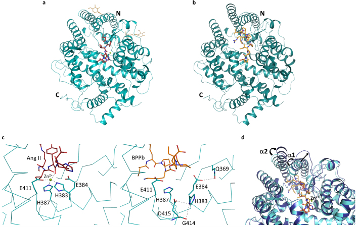
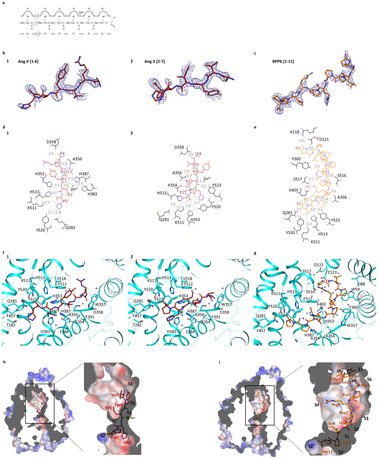
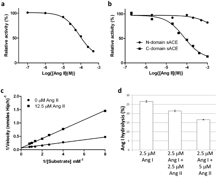
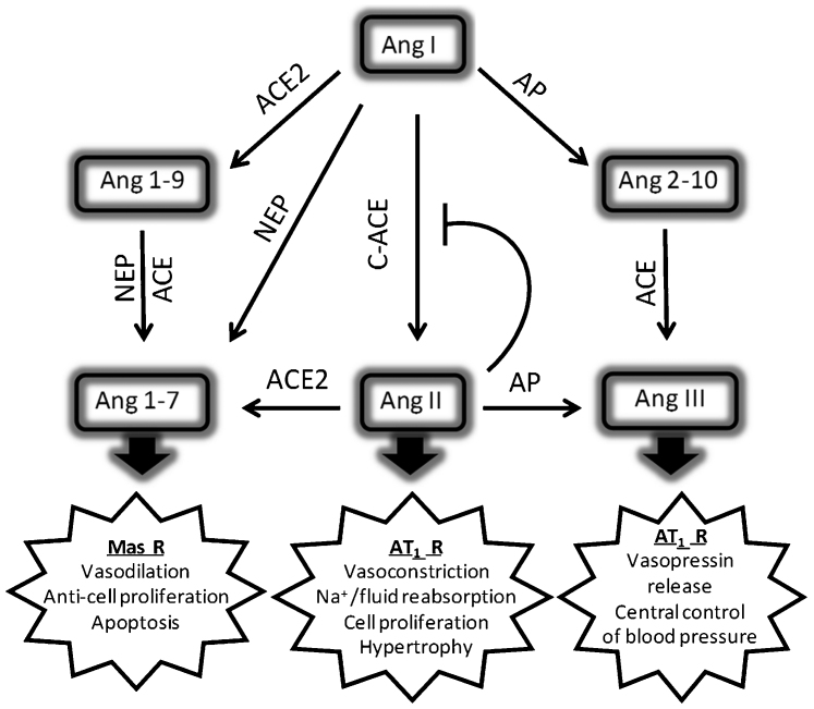
Angiotensin-I converting enzyme (ACE), a two-domain dipeptidylcarboxypeptidase, is a key regulator of blood pressure as a result of its critical role in the renin-angiotensin-aldosterone and kallikrein-kinin systems. Hence it is an important drug target in the treatment of cardiovascular diseases. ACE is primarily known for its ability to cleave angiotensin I (Ang I) to the vasoactive octapeptide angiotensin II (Ang II), but is also able to cleave a number of other substrates including the vasodilator bradykinin and N-acetyl-Ser-Asp-Lys-Pro (Ac-SDKP), a physiological modulator of hematopoiesis. For the first time we provide a detailed biochemical and structural basis for the domain selectivity of the natural peptide inhibitors of ACE, bradykinin potentiating peptide b and Ang II. Moreover, Ang II showed selective competitive inhibition of the carboxy-terminal domain of human somatic ACE providing evidence for a regulatory role in the human renin-angiotensin system (RAS).


Figures

Figure 1
Figure 1
(a) Substrate-bound human C-domain sACE crystal structure.C-domain ACE (cyan) in cartoon representation, with Ang II in red sticks, glycosylated carbohydrates in yellow sticks. The catalytic zinc ion is shown as a green sphere. (b) BPPb- bound C-domain ACE crystal structure. C-domain ACE (cyan) in cartoon representation, with BPPb in orange sticks. (c) Loss of the zinc ion coordination upon C-domain ACE binding to BPPb. Left panel, zinc coordination in the Ang II-bound C-domain ACE, with a classical tetrahedral motif, bound zinc ion as green sphere. Right panel, movement of the coordinating His383. (d) Conformational changes of C-domain ACE upon BPPb binding. Ang II and BPPb-bound C-domain ACE (cyan and blue respectively) in cartoon representation. Ang II and BPPb are shown as as red and orange sticks respectively. The zinc ion is only present in the Ang II-bound structure. Movement of the α1 and α2 helices is highlighted.
Figure 2
Figure 2
(a) Schematic model of the ACE active site.The relative positions of the enzyme subsites (Sn, S3, S2, S1, S1′, S2′), the residues of peptide inhibitors (Pn, P3, P2, P1, P1′, P2′) and the position of the catalytic zinc ion are shown. (b) Portions of the difference (Fo-Fc) electron density map for the bound Ang II peptide. Electron density map is contoured at 2.5σ level. The picture was created using a Fourier difference density map in which the peptide atoms were omitted. The two figures represent the interpreted structures of Ang II in two different (sliding) positions with 0.5 occupancy each for Ang II [1-6] and Ang II [2-7] respectively. (c) Portion of the difference (Fo-Fc) electron density map for the BPPb peptide (contoured at 2.5σ level). The picture was created from a Fourier difference density map in which the peptide atoms were omitted. (d) Detailed schematic of Ang II binding to C-domain ACE. Hydrogen bonds are highlighted (dashed lines) with Ang II in red and panel 1 with Ang II [1–6] and 2 with Ang II [2–7]. (e) Schematic diagram of BPPb binding to C-domain ACE. Hydrogen bonds are highlighted (dashed lines) with BPPb in orange. (f) Detailed binding of Ang II with the C-domain ACE. ACE in cartoon representation (cyan) with residues involved in binding highlighted (sticks). Ang II in red sticks, zinc ion as green sphere and water molecules in light blue are shown. Panel 1 with Ang II [1–6] and 2 with Ang II [2–7]. (g) Detailed binding of BPPb to C-domain ACE. ACE in cartoon representation (cyan) with residues involved in binding highlighted (sticks). BPPb in orange sticks, zinc ion as green sphere and water molecules in light blue. (h) Surface representation of Ang II binding (in red sticks) at the active site with electrostatic potential (red, blue for negative and positive potential respectively) computed with the APBS tool in PyMOL. The catalytic zinc ion is shown as a green sphere. (i) Surface representation of BPPb binding (in orange sticks) at the active site drawn as described in (h).
Figure 3
Figure 3
(a) Inhibition of human sACE by Ang II.Serum was used as the source of soluble sACE. ACE activity was assayed using HHL as substrate in presence of various concentrations of Ang II as described in the Methods. Data is expressed as a percentage of uninhibited activity and each data point is the mean of three replicates. A Hill slope of 1 was obtained using GraphPad© , which is consistent with simple linear competitive inhibition. (b) Inhibition of human N- and C-domain ACE by Ang II. ACE activity of recombinant N-domain and C-domain was determined using HHL as substrate in presence of various concentrations of Ang II as described in the Supplementary material. Data is expressed as a percentage of uninhibited activity and each data point is the mean of three replicates. A Hill slope of 1 for the inhibition of C-domain ACE was obtained using GraphPad© , which is consistent with simple linear competitive inhibition. (c) Lineweaver-Burk plot showing the competitive nature of the inhibition by Ang II of the hydrolysis of HHL by C-domain ACE. (d) Ang II inhibits Ang I conversion by human sACE. The impact of Ang II (2.5 and 5 µM) on the conversion of Ang I (2.5 µM) to Ang II by human sACE (serum) was determined by quantifying the decline in Ang I using HPLC. The results are expressed as % hydrolysis and are the means ± SEM (n = 4). HPLC analysis showed that Ang II was metabolically stable in serum under the same conditions as used for the inhibitor studies.
Figure 4
Figure 4. Schematic illustrating the roles of aminopeptidase (AP), carboxypeptidases (ACE and ACE2) and neprilysin (NEP) in the metabolism of angiotensin peptides.
Ang 1–7 (Asp-Arg-Val-Tyr-Ile-His-Pro), which can be formed either by the carboxypeptidase activity of ACE2 on Ang II (Asp-Arg-Val-Tyr-Ile-His-Pro-Phe) or by endopeptidase cleavage of Ang I (Asp-Arg-Val-Tyr-Ile-His-Pro-Phe-His-Leu) by neprilysin, antagonises the negative actions of Ang II on cardiovascular and renal physiology. The opposing effects of Ang II and Ang 1–7 are carried out primarily via the AT1 and Mas receptors (R), respectively. Ang III (Arg-Val-Tyr-Ile-His-Pro-Phe) is the principal active peptide in the brain RAS and its formation is regulated by aminopeptidase A. Homeostatic regulation of Ang I/Ang II levels in the kidney or heart through product inhibition of C-domain ACE might not only be important for regulating direct actions of the peptide, but also for the formation of Ang 1–7 and Ang 2–10 from Ang I by NEP and aminopeptidase A, respectively . The C-domain of sACE is primarily responsible for the formation of Ang II, but the relative contributions of the two domains of sACE to the hydrolysis of Ang 2–10 and Ang 1–9 are not known.

References

    1. Bader M. Tissue renin-angiotensin-aldosterone systems: Targets for pharmacological therapy. Annu. Rev. Pharmacol. Toxicol. 50, 439–465 (2010). - PubMed
    1. Kumar R., Singh V. P. & Baker K. M. The intracellular renin-angiotensin system: a new paradigm. Trends Endocrinol. Metab. 18, 208–214 (2007). - PubMed
    1. Corvol P., Eyries M. & Soubrier F. in Handbook of Proteolytic Enzymes edited by A. J. Barrett, N. D. Rawlings and J. F. Woessner (Elsevier Academic Press, Amsterdam, 2004), Vol. 1, pp. 332–346.
    1. Soffer R. L. Angiotensin-converting enzyme and the regulation of vasoactive peptides. Annu. Rev. Biochem. 45, 73–94. (1976). - PubMed
    1. Paul M., Poyan Mehr A. & Kreutz R. Physiology of local renin-angiotensin systems. Physiol. Rev. 86, 747–803 (2006). - PubMed

Publication types

MeSH terms