. 2012:2012:395329.
doi: 10.1155/2012/395329. Epub 2012 Sep 27.

H11/HspB8 and Its Herpes Simplex Virus Type 2 Homologue ICP10PK Share Functions That Regulate Cell Life/Death Decisions and Human Disease

Affiliations

H11/HspB8 and Its Herpes Simplex Virus Type 2 Homologue ICP10PK Share Functions That Regulate Cell Life/Death Decisions and Human Disease

Laure Aurelian et al. Autoimmune Dis. 2012.

Abstract

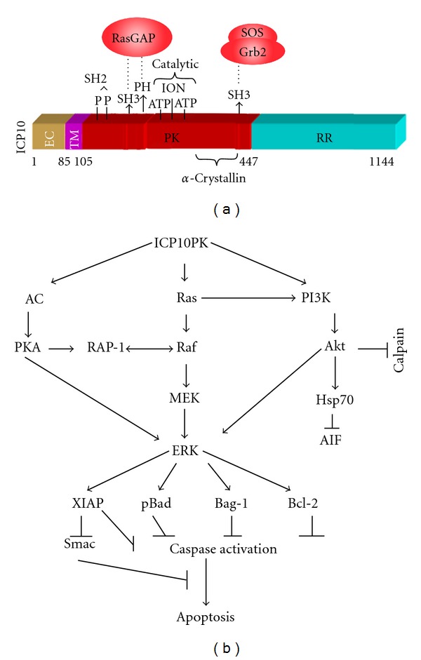
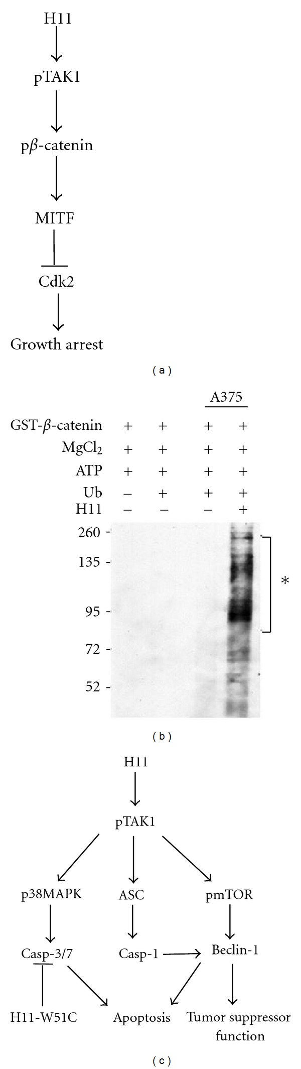
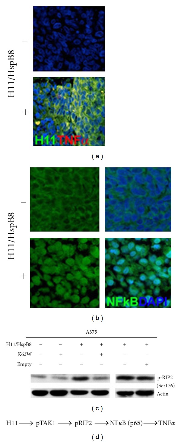
Small heat shock proteins (sHsp) also known as HspB are a large family of widely expressed proteins that contain a 90 residues domain known as α-crystallin. Here, we focus on the family member H11/HspB8 and its herpes simplex virus type 2 (HSV-2) homologue ICP10PK, and discuss the possible impact of this relationship on human disease. H11/HspB8 and ICP10PK are atypical protein kinases. They share multi-functional activity that encompasses signaling, unfolded protein response (UPR) and the regulation of life cycle potential. In melanocytes H11/HspB8 causes growth arrest. It is silenced in a high proportion of melanoma prostate cancer, Ewing's sarcoma and hematologic malignancies through aberrant DNA methylation. Its restored expression induces cell death and inhibits tumor growth in xenograft models, identifying H11/HspB8 as a tumor suppressor. This function involves the activation of multiple and distinct death pathways, all of which initiate with H11/HspB8-mediated phosphorylation of transforming growth factor β-activated kinase 1 (TAK1). Both ICP10PK and H11/HspB8 were implicated in inflammatory processes that involve dendritic cells activation through Toll-like receptor-dependent pathways and may contribute to the onset of autoimmunity. The potential evolutionary relationship of H11/HspB8 to ICP10PK, its impact on human disorders and the development of therapeutic strategies are discussed.


Figures

Figure 1
Figure 1
ICP10PK activates survival/proliferative pathways that initiate with Ras activation. (a) Schematic representation of the ICP10PK functional motifs. Shown are the binding sites for the Grb2-Sos complex and Ras-GAP, the PK catalytic motifs the TM and extracellular (EC) domains, and the α-crystallin domain. (b) The activated signaling pathways include the Ras/c-Raf/MEK/ERK, the PI3-K/Akt, and the AC/PKA. The pathways converge on ERK activation and modulate apoptosis regulatory proteins that control caspase activation (Bcl2 family) and caspase activity (iAP family) and their inhibitors (Smac). ICP10PK also inhibits calpain activation and AIF release. Ras activation is through recruitment of the GEF Sos and inhibition of the Ras-GAP through direct phosphorylation [–13].
Figure 2
Figure 2
The ICP10PK vector ΔRR inhibits UPR. (a) mSOD1 (G93A) transgenic rats were treated with ΔRR (107 pfu; 100 μL) or an equal volume of PBS in both hind limbs by intramuscular injection beginning at age of 85 days. The ΔRR-treated G93A rats lived significantly longer than their PBS-treated counterparts with a median survival of 195 and 154 days, respectively (P < 0.001 by log-rank Mantel-Cox analysis). Serial sections from the L4-L5 region collected on day 153 were stained in double immunofluorescence with antibodies to human SOD1 (Alexa Fluor 488 conjugated secondary antibody; green) and CHOP (Alexa Fluor 594 conjugated secondary antibody; red). DAPI nuclear staining is blue. Only the PBS treated animals showed evidence of aggregates containing CHOP and mSOD1 co-localization. Scale bar = 10 μm. (b). Neuronally differentiated N2a cells that were stably transfected with another mSOD1 mutant (G85R) were mock infected with PBS or infected with ΔRR or a vector that lacks ICP10PK (ΔPK) and treated with H2O2 (100 μM; 2 hrs) to induce oxidative stress. N2a cells stably transfected with the wt SOD1 were studied in parallel and served as control. At 24 hrs after infection the cells were stained by double immunofluorescence with Alexa Fluor 594 conjugated SOD1 antibody (red) and Alexa Fluor 488 conjugated pp38MAPK antibody (green). DAPI nuclear stain is in blue. Scale bar = 10 μm. Aggregates are not seen in the ΔRR-treated cells, associated with the inhibition of p38MAPK phosphorylation (activation).
Figure 3
Figure 3
H11/HsPB8 causes growth arrest and melanoma cell death through distinct TAK1-regulated pathways. (a) Schematic representation of H11/HspB8-induced growth arrest involves β-catenin phosphorylation by the activated TAK1. (b) GST-β-catenin phosphorylated when mixed with cell extracts containing H11/HspB8-activated TAK1 is ubiquitylated causing its degradation (∗). Ubiquitylation is not seen when GST-β-catenin is mixed with cell extracts that lack H11/HspB8 and therefore pTAK1. (c) H11/HspB8 induces different death pathways in the genetically diverse melanoma cells A375 and A2058 that initiate with TAK1 activation. In A375 cells, the TAK1/p38MAPK pathway activates caspase-3/7 to cause apoptosis. In A2058 cells, the TAK1/p38MAPK pathway activates caspase-3, but TAK1 also activates caspase-1 through ASC upregulation and upregulates Beclin-1 through mTOR phosphorylation at S2481 (pmTORS2481). Caspase-1 cleaves Beclin-1 to promote apoptosis, but Beclin-1 also contributes to cell death through still unknown tumor suppressor functions. The W51C mutant of H11/HspB8 has dominant-negative activity. It inhibits the death-inducing potential of the wild-type H11/HspB8 by inhibiting caspase activation through the activation of the B-Raf/MEK/ERK pathway [33].
Figure 4
Figure 4
H11/HspB8 induces TAK1-dependent TNF-α expression. (a) Serial sections from A2058 and A375 xenografts collected at the end of the follow-up period from mice subjected or not to restored H11/HspB8 expression were stained with antibodies to H11/HspB8 (Alexa Fluor 488 conjugated secondary antibody; green) and TNF-α (Alexa Fluor 594 conjugated secondary antibody; red). (b). Serial sections from the A2058 and A375 xenografts as in (a) were stained with Alexa Fluor 488 conjugated NF-κB p65 antibody. Restored H1/HspB8 activates NF-κB (intranuclear localization). (c) A375 cells stably transfected with tet-inducible H11/HspB8 were given Dox to restore H11/HspB8 expression alone or with the TAK1 dominant-negative mutant K63W or empty vector. Extracts collected 3 days later were immunoblotted with antibody to pRIP2 (Ser176). They were stripped and reprobed with antibody to actin (loading control). (d) Schematic representation of the pathways involved in H11/HspB8-induced TNF-α induction.

References

    1. Kappé G, Boelens WC, de Jong WW. Why proteins without an α-crystallin domain should not be included in the human small heat shock protein family HSPB. Cell Stress and Chaperones. 2010;15(4):457–461. - PMC - PubMed
    1. Hartl FU. Chaperone-assisted protein folding: the path to discovery from a personal perspective. Nature Medicine. 2011;17:1206–1210. - PubMed
    1. Mchaourab HS, Godar JA, Stewart PL. Structure and mechanism of protein stability sensors: chaperone activity of small heat shock proteins. Biochemistry. 2009;48(18):3828–3837. - PMC - PubMed
    1. Mymrikov EV, Seit-Nebi AS, Gusev NB. Large potentials of small heat shock proteins. Physiological Reviews. 2011;91:1123–1159. - PubMed
    1. Acunzo J, Katsogiannou M, Rocchi P. Small heat shock proteins HSP27 (HspB1), αB-crystallin (HspB5) and HSP22 (HspB8) as regulators of cell death. International Journal of Biochemistry & Cell Biology. 2012;44(10):1622–1631. - PubMed

LinkOut - more resources