. 2012 Oct 11:7:78.
doi: 10.1186/1750-1172-7-78.

Quantitative analysis of ciliary beating in primary ciliary dyskinesia: a pilot study

Affiliations

Quantitative analysis of ciliary beating in primary ciliary dyskinesia: a pilot study

Jean-François Papon et al. Orphanet J Rare Dis. .

Abstract

Background: Primary ciliary dyskinesia (PCD) is a rare congenital respiratory disorder characterized by abnormal ciliary motility leading to chronic airway infections. Qualitative evaluation of ciliary beat pattern based on digital high-speed videomicroscopy analysis has been proposed in the diagnosis process of PCD. Although this evaluation is easy in typical cases, it becomes difficult when ciliary beating is partially maintained. We postulated that a quantitative analysis of beat pattern would improve PCD diagnosis. We compared quantitative parameters with the qualitative evaluation of ciliary beat pattern in patients in whom the diagnosis of PCD was confirmed or excluded.

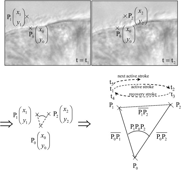
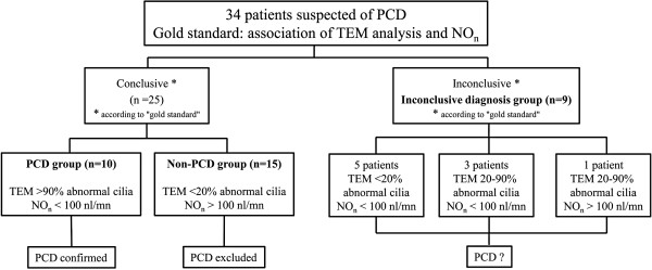
Methods: Nasal nitric oxide measurement, nasal brushings and biopsies were performed prospectively in 34 patients with suspected PCD. In combination with qualitative analysis, 12 quantitative parameters of ciliary beat pattern were determined on high-speed videomicroscopy recordings of beating ciliated edges. The combination of ciliary ultrastructural abnormalities on transmission electron microscopy analysis with low nasal nitric oxide levels was the "gold standard" used to establish the diagnosis of PCD.

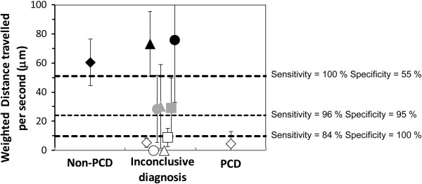
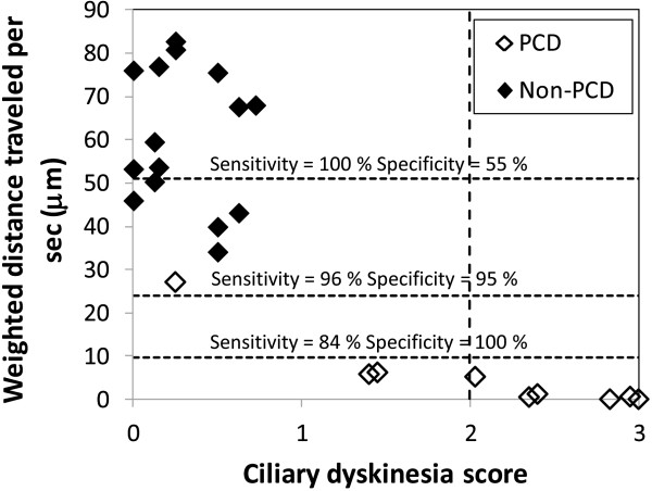
Results: This "gold standard" excluded PCD in 15 patients (non-PCD patients), confirmed PCD in 10 patients (PCD patients) and was inconclusive in 9 patients. Among the 12 parameters, the distance traveled by the cilium tip weighted by the percentage of beating ciliated edges presented 96% sensitivity and 95% specificity. Qualitative evaluation and quantitative analysis were concordant in non-PCD patients. In 9/10 PCD patients, quantitative analysis was concordant with the "gold standard", while the qualitative evaluation was discordant with the "gold standard" in 3/10 cases. Among the patients with an inconclusive "gold standard", the use of quantitative parameters supported PCD diagnosis in 4/9 patients (confirmed by the identification of disease-causing mutations in one patient) and PCD exclusion in 2/9 patients.

Conclusions: When the beat pattern is normal or virtually immotile, the qualitative evaluation is adequate to study ciliary beating in patients suspected for PCD. However, when cilia are still beating but with moderate alterations (more than 40% of patients suspected for PCD), quantitative analysis is required to precise the diagnosis and can be proposed to select patients eligible for TEM.


Figures

Figure 1
Figure 1
Determination of digital high-speed videomicroscopy parameters of ciliary beat pattern of a cilium. The three positions of the cilium (base of the cilium, P0, positions of the tip before the active and recovery strokes, P1 and P2 respectively) and the five time-points of beating (start and arrival of the active stroke, t1 and t2 respectively, start and arrival of the recovery stroke, t3 and t4 respectively, and start of the following cycle, t5) were measured. Twelve parameters were then determined from these measurements:. * The cilia length: Lc=maxP0P¯1,P0P2¯ where PiPj¯ designates the euclidean distance between the points Pi and Pj. In cartesian coordinates PiPj¯=xi-xj2+yi-yj2 *The angle of beating, P1P0P2^, is given by the law of cosines: P1P0P2^=ArcosP0P¯12+P0P2¯2-P1P2¯22P0P¯1P0P2¯ *The global frequency: Fg=1t5-t1 *The power stroke duration: Dp=(t2 - t1) *The recovery duration: Dr=(t4 - t3) *The pause after stroke: Ps=t3 - t2 *The pause after recovery: Pr=t5 - t4 *The total pause: Pt=Ps+Pr *The distance travelled per second = LcP1P0P2^fg1second *The area swept per second =P1P0P2^/2Lc2fg1second *The weighted distance travelled per second = % of beating edgesLcP1P0P2^fg1second *The weighted area swept per second = % of beating edgesP1P0P2^/2Lc2fg1second.
Figure 2
Figure 2
Determination of the patient groups according to the “gold standard”.
Figure 3
Figure 3
Comparison of digital high-speed videomicroscopy parameters in the non-PCD and PCD groups. The area under the curve (AUC) of the ROC curve was ≥ 0.69 for each parameter. The distance travelled per second (by the cilium tip) and the area swept per second (by the cilium), weighted by the percentage of beating ciliated edges, presented the best AUC (0.984 and 0.977, respectively).
Figure 4
Figure 4
Evaluation of the efficiency of digital high-speed videomicroscopy in nine patients with an inconclusive diagnosis. In this group, the mean distance travelled by the cilium tip per second weighted by the percentage of beating ciliated edges, was used to discriminate patients. The PCD and non-PCD cutoffs were < 10 μm and > 51 μm respectively. The symbols of the non-PCD and PCD groups represent the mean values ± standard deviation. In the inconclusive group, white symbols represent patients in whom PCD was possibly confirmed, black symbols represent patients in whom PCD was possibly excluded and grey symbols represent patients in whom digital high-speed videomicroscopy was not helpful for PCD diagnosis.
Figure 5
Figure 5
Quantitative analysis versus qualitative evaluation. The results of the weighted distance travelled per second is plotted versus the dyskinesia score in the patients with conclusive gold standard. According to Stannard et al. [13], a score ≥ 2 was described as the best predictor of PCD.

References

    1. Bush A, Chodhari R, Collins N, Copeland F, Hall P, Harcourt J, Hariri M, Hogg C, Lucas J, Mitchison HM. et al.Primary ciliary dyskinesia: current state of the art. Arch Dis Child. 2007;92:1136–1140. doi: 10.1136/adc.2006.096958. - DOI - PMC - PubMed
    1. Afzelius BA. The immotile-cilia syndrome: a microtubule-associated defect. CRC Crit Rev Biochem. 1985;19:63–87. doi: 10.3109/10409238509086788. - DOI - PubMed
    1. Barbato A, Frischer T, Kuehni CE, Snijders D, Azevedo I, Baktai G, Bartoloni L, Eber E, Escribano A, Haarman E. et al.Primary ciliary dyskinesia: a consensus statement on diagnostic and treatment approaches in children. Eur Respir J. 2009;34:1264–1276. doi: 10.1183/09031936.00176608. - DOI - PubMed
    1. Coren ME, Meeks M, Morrison I, Buchdahl RM, Bush A. Primary ciliary dyskinesia: age at diagnosis and symptom history. Acta Paediatr. 2002;91:667–669. doi: 10.1111/j.1651-2227.2002.tb03299.x. - DOI - PubMed
    1. Leigh MW, Pittman JE, Carson JL, Ferkol TW, Dell SD, Davis SD, Knowles MR, Zariwala MA. Clinical and genetic aspects of primary ciliary dyskinesia/Kartagener syndrome. Genet Med. 2009;11:473–487. doi: 10.1097/GIM.0b013e3181a53562. - DOI - PMC - PubMed

Publication types

LinkOut - more resources