. 2012 Oct 30;51(43):8563-70.
doi: 10.1021/bi3011797. Epub 2012 Oct 18.

A bacterial hemerythrin domain regulates the activity of a Vibrio cholerae diguanylate cyclase

Affiliations

A bacterial hemerythrin domain regulates the activity of a Vibrio cholerae diguanylate cyclase

Ruth A Schaller et al. Biochemistry. .

Abstract

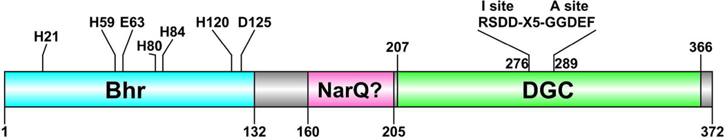
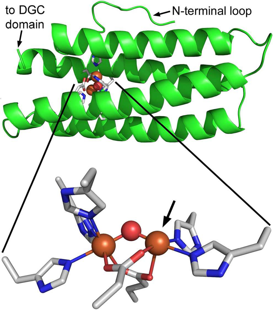
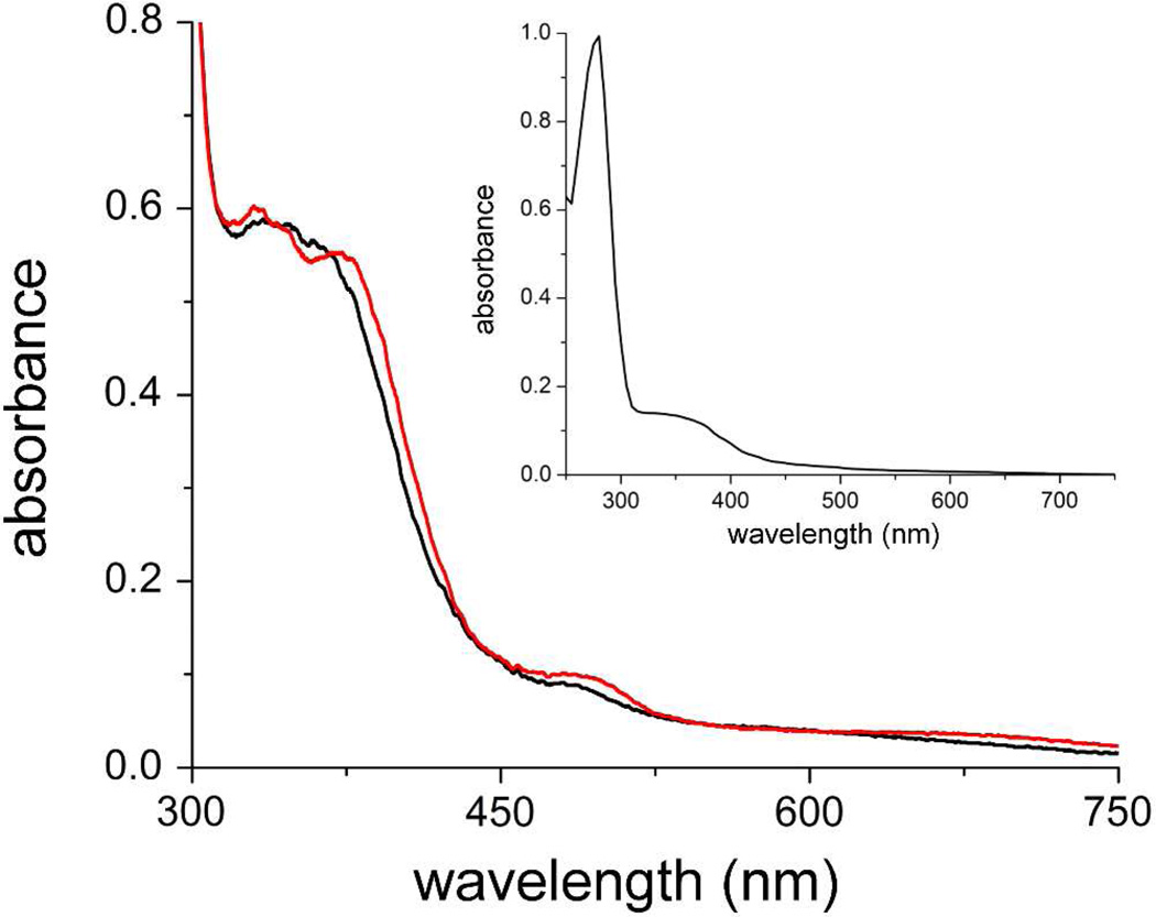
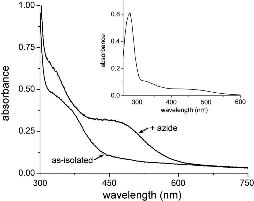
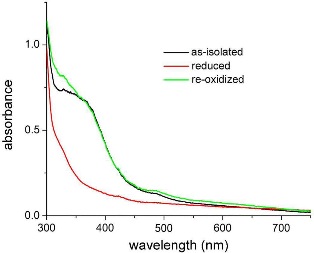
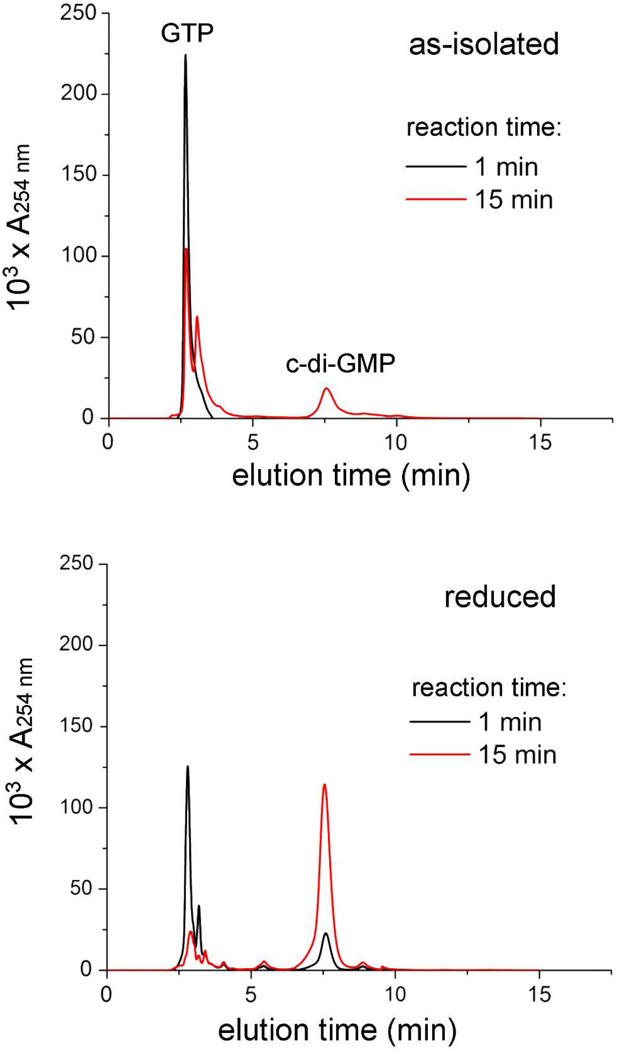
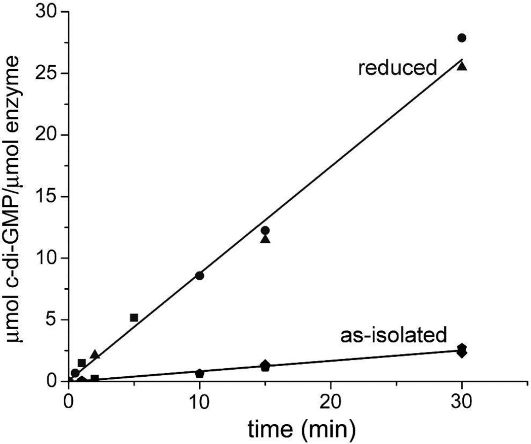
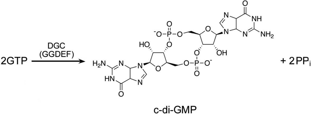
The first demonstrated example of a regulatory function for a bacterial hemerythrin (Bhr) domain is reported. Bhrs have a characteristic sequence motif providing ligand residues for a type of non-heme diiron site that is known to bind O(2) and undergo autoxidation. The amino acid sequence encoded by the VC1216 gene from Vibrio cholerae O1 biovar El Tor str. N16961 contains an N-terminal Bhr domain connected to a C-terminal domain characteristic of bacterial diguanylate cyclases (DGCs) that catalyze formation of cyclic di-(3',5')-guanosine monophosphate (c-di-GMP) from GTP. This protein, Vc Bhr-DGC, was found to contain two tightly bound non-heme iron atoms per protein monomer. The as-isolated protein showed the spectroscopic signatures of oxo/dicarboxylato-bridged non-heme diferric sites of previously characterized Bhr domains. The diiron site was capable of cycling between diferric and diferrous forms, the latter of which was stable only under anaerobic conditions, undergoing rapid autoxidation upon being exposed to air. Vc Bhr-DGC showed approximately 10 times higher DGC activity in the diferrous than in the diferric form. The level of intracellular c-di-GMP is known to regulate biofilm formation in V. cholerae. The higher DGC activity of the diferrous Vc Bhr-DGC is consistent with induction of biofilm formation in low-dioxygen environments. The non-heme diiron cofactor in the Bhr domain thus represents an alternative to heme or flavin for redox and/or diatomic gas sensing and regulation of DGC activity.


Figures

Figure 1
Figure 1
Annotated domains and sequence motifs for the 372-residue Vc Bhr-DGC encoded by VC1216. Predicted iron ligand residues are indicated in the Bhr domain. Predicted active (A site) and inhibitory (I site) sequence motifs are indicated in the DGC domain. NarQ? refers to provisional annotation as homologous to sequences in nitrate/nitrite sensor proteins. The residue-132 C-terminal limit of the Bhr domain is from Bailly et al. Sequence limits for the other domains are those listed at http://www.ncbi.nlm.nih.gov/protein/15641229?report=graph.
Figure 2
Figure 2
Homology structural model of the Bhr domain (residues 3–133) in Vc Bhr-DGC generated using the DcrH-Hr diferric-azido structure (PDB entry 2avk) as template. The modeled protein backbone is shown in green cartoon mode, and modeled iron ligand side chains in CPK-colored stick mode. Iron atoms and bridging oxo from 2avk coordinates are shown as orange and red spheres, respectively. The azide atoms are omitted for clarity. Arrow indicates the azide coordination position. Images were generated in PyMOL (Delano Scientific LLC).
Figure 3
Figure 3
UV-vis absorption spectra of ~90 µM as-isolated Vc Bhr-DGC in 250 mM NaCl 125 mM imidazole in 50 mM MOPS + 20% (v/v) glycerol pH 7.3 (red trace) or after dialysis against 50 mM MOP + 20% (v/v) glycerol pH 7.3 to remove NaCl and imidazole (black trace). Inset shows the spectrum of the dialyzed protein including the 280-nm region.
Figure 4
Figure 4
UV-vis absorption spectra of as-isolated Vc Bhr-DGC in 50 mM MOPS, 10% glycerol pH 7.3 and after addition of sodium azide to a final concentration of 50 mM. Inset shows the spectrum of the protein + azide including the 280 nm region. The azide-containing sample was incubated at 4 °C for 4 hours prior to recording the spectrum.
Figure 5
Figure 5
UV-vis absorption spectra obtained upon redox cycling of Vc Bhr-DGC in 50 mM MOPS, 10% glycerol pH 7.3. A solution of the as-isolated protein (black trace) was reduced anaerobically with ~1 equivalent of sodium dithionite, then incubated for three hours anaerobically before recording the spectrum (red trace). This reduced sample was then re-oxidized by exposure to air, and the spectrum was recorded again (green trace).
Figure 6
Figure 6
Congo Red plate assay for DGC activity. Each quadrant of the LB/ampicillin/±IPTG agar plates was spotted with a 100-microliter aliquot of cultures of E. coli BL21(DE3) that had been transformed with either the parent expression plasmid, pAG8H, (upper two quadrants, labeled “empty vector”), or pAG8H containing the Vc Bhr-DGC gene (lower two quadrants, labeled “+VC1216”). The plates were then incubated for 72 hrs at 37 °C. Colonies showing no visible Congo Red stain in this view are circled. The circled colonies are better visualized in the magnified image (Figure S6).
Figure 7
Figure 7
HPLC traces showing production of c-di-GMP and consumption of GTP. Assay and HPLC and conditions are described in Materials and Methods. The Vc Bhr-DGC concentrations were 74 µM and 64 µM (monomer basis) for the as-isolated and reduced proteins, respectively.
Figure 8
Figure 8
Rates of c-di-GMP production for as-isolated and dithionite-reduced Vc Bhr-DGC. Assay conditions are described in Materials and Methods. Micromolar enzyme concentrations (protein monomer basis) are for reduced: 6.25 (circles), 10 (triangles), and 64 (squares); for as-isolated: 6.25 (diamonds), 74 (pentagons) and 100 (squares).
Scheme 1
Scheme 1

Similar articles

Cited by

References

    1. Romling U, Gomelsky M, Galperin MY. C-di-GMP: the dawning of a novel bacterial signalling system. Mol. Microbiol. 2005;57:629–639. - PubMed
    1. Ryan RP, Fouhy Y, Lucey JF, Dow JM. Cyclic di-GMP signaling in bacteria: recent advances and new puzzles. J. Bacteriol. 2006;188:8327–8334. - PMC - PubMed
    1. Hengge R. Principles of c-di-GMP signalling in bacteria. Nature Rev. Microbiol. 2009;7:263–273. - PubMed
    1. Wolfe AJ, Visick KL, editors. The second messenger cyclic di-GMP. Washington, DC: ASM Press; 2010.
    1. Sondermann H, Shikuma NJ, Yildiz FH. You've come a long way: c-di- GMP signaling. Curr Opin Microbiol. 2012;15:140–146. - PMC - PubMed

Publication types

MeSH terms

LinkOut - more resources