Review
. 2012:28:719-41.
doi: 10.1146/annurev-cellbio-101011-155739.

Cardiac regenerative capacity and mechanisms

Affiliations
Review

Cardiac regenerative capacity and mechanisms

Kazu Kikuchi et al. Annu Rev Cell Dev Biol. 2012.

Abstract

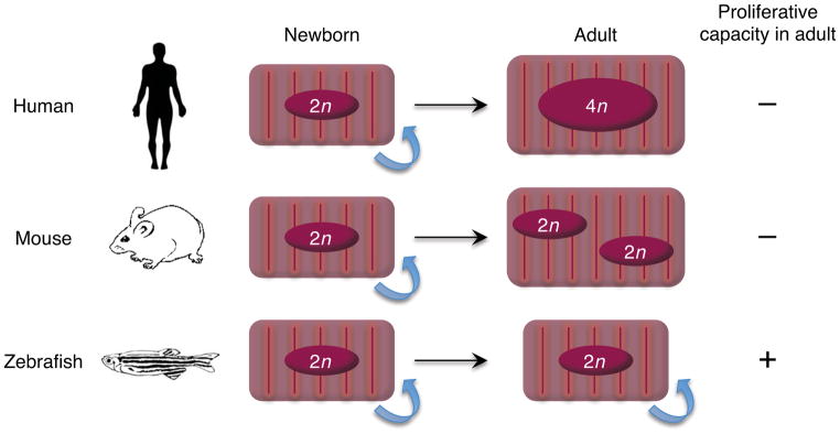
The heart holds the monumental yet monotonous task of maintaining circulation. Although cardiac function is critical to other organs and to life itself, mammals are not equipped with significant natural capacity to replace heart muscle that has been lost by injury. This deficiency plays a role in leaving millions worldwide vulnerable to heart failure each year. By contrast, certain other vertebrate species such as zebrafish are strikingly good at heart regeneration. A cellular and molecular understanding of endogenous regenerative mechanisms and advances in methodology to transplant cells together project a future in which cardiac muscle regeneration can be therapeutically stimulated in injured human hearts. This review focuses on what has been discovered recently about cardiac regenerative capacity and how natural mechanisms of heart regeneration in model systems are stimulated and maintained.


Figures

Figure 1
Figure 1
Nuclear dynamics and proliferative capacity of cardiomyocytes during growth. Cardiomyocytes in fetal humans and mice typically have a single nucleus with a diploid genome (2n) and increase mass through cell division. Human cardiomyocytes can proliferate for the first few months after birth, but are believed to lose this capacity early in life. These cells typically undergo rounds of DNA replication without karyokinesis or cytokinesis, resulting largely in mononucleated cardiomyocytes with tetraploid (4n) or higher DNA content. Murine cardiomyocytes can divide robustly until the first few days after birth, after which the majority withdraw from the cell cycle. These cells undergo additional DNA replication with karyokinesis but not cytokineis, resulting in binucleated cardiomyocytes that are diploid (2n) in each nucleus (Laflamme & Murry 2011). By contrast, most cardiomyocytes in zebrafish hearts are mononucleated with a diploid genome (2n) throughout life, with significant proliferative capacity (Wills et al 2007).
Figure 2
Figure 2
Endogenous cardiac stem or progenitor cells in the postnatal mammalian heart. These cell populations have been directly identified based on cell surface marker expression (c-Kit+ and Sca-1+ cells), genetic marker expression (Islet1+ cells), and dye-efflux function (SP cells), or indirectly identified as cell clusters derived from the culture of dissociated cardiac tissues (CSs and cCFU-Fs).
Figure 3
Figure 3
Cellular origins of regenerating cardiomyocytes. (a) Genetic fate-mapping to determine the source of new cardiomyocytes during zebrafish heart regeneration. Treatment with tamoxifen leads to marking of nearly all cardiomyocytes with EGFP. Following resection, new cardiomyocytes expressed EGFP, indicating derivation from existing cardiomyocytes rather than a different unmarked cell source (Kikuchi et al 2010). (b) A similar fate-mapping experiment was performed in the neonatal mouse heart, in this case with an inducible LacZ reporter. Although the labeling of cardiomyocytes with this system was ~60%, the ratio of labeled cells was maintained between the regenerate and uninjured myocardium. This result indicates that the new cardiomyocytes are derived from existing cardiomyocytes rather than a progenitor cell (Porrello et al 2011b). (c) Fate-mapping experiments were performed in the adult mouse heart with an inducible GFP reporter. The labeling of cardiomyocytes with this system was ~83%, and the ratio of labeled cells was decreased to ~68% in areas bordering the MI, and decreased to ~76% in hearts subjected to pressure overload. Thus, stem or progenitor cells might refresh cardiomyocytes in hearts after pressure overload or myocardial infarction (Hsieh et al 2007).
Figure 4
Figure 4
Potential role of Thymosin β4 in cardiomyocyte differentiation from non- myocytes. In adult mouse hearts, Wt1 is absent or expressed at low levels in epicardial cells. Epicardial cells re-express Wt1 after infarction, proliferate, and give rise to mesenchymal cells expressing smooth muscle and fibroblast cell markers, but never differentiate into cardiomyocytes (Zhou et al 2011). With Thymosin β4 injections prior to the injury, more epicardial cells induce Wt1 expression after MI, some of which coexpress cardiac progenitor markers such as Islet1. Evidence suggests that some of these activated epicardial cells transdifferentiate to cardiomyocytes (Smart et al 2011).
Figure 5
Figure 5
Injury responses of the three major cardiac cell types in zebrafish. (a) Ostensibly the entire epicardium activates developmental genes such as tbx18 and raldh2 (violet) within several days following injury. Enhanced gene expression localizes to the wound area by two weeks post-injury (arrowheads). (b) Endocardial cells in the entire heart upregulate raldh2 (violet) within hours of amputation, before localization to the injury site. (c) Myocardial activation, represented by induction of a gata4:EGFP reporter, is first detected throughout the compact layer of the myocardium by 7 days post-amputation (dpa) and becomes localized to the regenerating myocardium by 14–30 dpa (arrowheads). Cardiomyocyte nuclei are visualized by Mef2. ba, bulbus arteriosus; at, atrium; ve, ventricle. Dotted lines indicate approximate amputation planes. Modified from previous studies (Kikuchi et al 2011b, Kikuchi et al 2010, Lepilina et al 2006).

References

    1. Ambasudhan R, Talantova M, Coleman R, Yuan X, Zhu S, et al. Direct reprogramming of adult human fibroblasts to functional neurons under defined conditions. Cell Stem Cell. 2011;9:113–18. - PMC - PubMed
    1. Bader D, Oberpriller JO. Repair and reorganization of minced cardiac muscle in the adult newt (Notophthalmus viridescens) Journal of Morphology. 1978;155:349–57. - PubMed
    1. Balsam LB, Wagers AJ, Christensen JL, Kofidis T, Weissman IL, Robbins RC. Haematopoietic stem cells adopt mature haematopoietic fates in ischaemic myocardium. Nature. 2004;428:668–73. - PubMed
    1. Barker N, van Es JH, Kuipers J, Kujala P, van den Born M, et al. Identification of stem cells in small intestine and colon by marker gene Lgr5. Nature. 2007;449:1003–07. - PubMed
    1. Bearzi C, Rota M, Hosoda T, Tillmanns J, Nascimbene A, et al. Human cardiac stem cells. Proceedings of the National Academy of Sciences of the United States of America. 2007;104:14068–73. - PMC - PubMed

Publication types

LinkOut - more resources