Review
. 2012 Dec;37(12):526-33.
doi: 10.1016/j.tibs.2012.09.001. Epub 2012 Oct 8.

Membrane curvature and its generation by BAR proteins

Affiliations
Review

Membrane curvature and its generation by BAR proteins

Carsten Mim et al. Trends Biochem Sci. 2012 Dec.

Abstract

Membranes are flexible barriers that surround the cell and its compartments. To execute vital functions such as locomotion or receptor turnover, cells need to control the shapes of their membranes. In part, this control is achieved through membrane-bending proteins, such as the Bin/amphiphysin/Rvs (BAR) domain proteins. Many open questions remain about the mechanisms by which membrane-bending proteins function. Addressing this shortfall, recent structures of BAR protein:membrane complexes support existing mechanistic models, but also produced novel insights into how BAR domain proteins sense, stabilize, and generate curvature. Here we review these recent findings, focusing on how BAR proteins interact with the membrane, and how the resulting scaffold structures might aid the recruitment of other proteins to the sites where membranes are bent.


Figures

Figure 1
Figure 1. Curvature and membrane structures
(A) Membrane structures, like invaginations, are composed of areas with different curvatures. Note that the sign of the curvature depends on the vantage point; hereafter curvature will be referred to as seen from the cytosol. On the top is a schematic view of a plasma membrane invagination displaying various curvatures. The flat membrane has zero (0) curvature (yellow). At the base of the invagination (brown) the curvature is negative (−). The part of the membrane that reaches further into the cytosol possesses positive (+) curvature. The bottom shows a membrane tubule, which is a ubiquitous structure in cells (e.g. T-tubules in muscle cells). These structures are created and maintained by proteins (blue crescents). The curvature is positive along the circumference of the tubule and zero along the tubular axis. (C) Schematic view of the mechanisms by which BAR domains can generate curvature. Upper panel: the crescent shaped BAR domain interacts with the bilayer through electrostatic interactions, imposing its intrinsic shape onto the membrane. This mechanism is known as the ‘scaffolding mechanism’. Lower panel: alternatively, BAR domains can introduce an amphipathic structure, such as a helix, into one leaflet of the membrane. This ‘wedge’ displaces lipids, which will cause the membrane to bend towards the BAR-domain. This mechanism is referred to as the ‘wedging mechanism’.
Figure 2
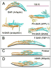
Figure 2. Structures of BAR domains from different subfamilies
All structures are depicted as dimers (cyan and orange) (scale bar 130Å). The black line underneath each structure was introduced to show the intrinsic curvature of each domain. (A) Left, above: side view of the BAR domain of arfaptin 1 (pdb ID 1I49). BAR domains exhibit the highest positive curvature within the BAR superfamily. Classic BAR domains are further subdivided based on additional membrane binding domains. For clarity, different BAR domains are shown as top view. The N-BAR domain (endophilin, pdb courtesy of G. Voth from [76], left, below) contains a N-terminal helix (yellow and green) and a second set of amphipathic helices known as ‘insert helices’ (orange and cyan). APPL1 (right, above) is an example of a PH-BAR domain that contains an additional pleckstrin-homology domain (PH) (pdb ID 2Z0O). Lastly, PX-BAR proteins (right, below), such as sorting nexin 9 (SNX9, pdb ID 2RAI), are comprised of a phox-homology domain (PX) and a BAR domain. (B) Sideview of the F-BAR domain from FBP (pdb ID 2EFL) exemplifies a shallow, positive curvature that is typical for this subfamily. (C) The Pinkbar is the only instance of a BAR protein that promotes zero curvature; its BAR domain shows no intrinsic curvature (pdb ID 3OK8). (D) I-BAR domain proteins are the only subfamily that can bend membranes to generate structures with negative curvature (IRSp53, pdb ID 1Y2O).
Figure 3
Figure 3. Structures of membrane-bound BAR domains
All structures are of protein-coated membrane tubules that have been reconstructed from electron microscopic images. The density of the protein is colored in silver, whereas the membrane is blue. (A) Structure the CIP4 F-BAR domain scaffold (pdb ID 2EFL) (EMDB accession code 1471). Top: side view of the membrane tubule. One copy of the F-BAR domain of the closely related FBP17 was fitted to highlight organization of the protein on the membrane (scale bar 220Å). F-BAR dimers form extensive lateral interactions that give rise to a stiff protein coat, which imposes its shape on the membrane. The cartoon highlights the high degree of overlap between adjacent F-BAR dimers. Middle: cross-section along the membrane tubule axis. Bottom: cross-section perpendicular to the membrane tubule axis. The orange bars demarcate the approximate boundaries of the membrane bilayer. (B) Structure of the endophilin N-BAR scaffold (EMDB accession code 5365). Top: side view of the membrane tubule. One dimer of the N-BAR domain (pdb ID 1ZWW) was fitted to show the density occupied by the protein (scale bar 130Å). Additionally, the amphipathic helices present in endophilin are depicted as cylinders for better visibility. N-BAR scaffolds are held together by interactions between the N-terminal H0 helices (see cartoon). Middle: cross-section along the membrane tubule axis, illustrating the positioning of the BAR-domain and the slope of the insert helices. Bottom: cross-section perpendicular to the membrane tubule axis. The leaflet closest to the N-BAR domain is continuous with the density of the protein, indicating a loss of order of the lipid head groups.

References

    1. Roth TF, Porter KR. Yolk Protein Uptake in the Oocyte of the Mosquito Aedes Aegypti. L. The Journal of cell biology. 1964;20:313–332. - PMC - PubMed
    1. Chen MS, et al. Multiple forms of dynamin are encoded by shibire, a Drosophila gene involved in endocytosis. Nature. 1991;351:583–586. - PubMed
    1. van der Bliek AM, Meyerowitz EM. Dynamin-like protein encoded by the Drosophila shibire gene associated with vesicular traffic. Nature. 1991;351:411–414. - PubMed
    1. Kosaka T, Ikeda K. Possible temperature-dependent blockage of synaptic vesicle recycling induced by a single gene mutation in Drosophila. Journal of neurobiology. 1983;14:207–225. - PubMed
    1. Takei K, et al. Functional partnership between amphiphysin and dynamin in clathrin-mediated endocytosis. Nature cell biology. 1999:33–39. - PubMed

MeSH terms

LinkOut - more resources