Review
. 2013 Jul:136:271-9.
doi: 10.1016/j.jsbmb.2012.09.021. Epub 2012 Oct 8.

Protective role of vitamin D signaling in skin cancer formation

Affiliations
Review

Protective role of vitamin D signaling in skin cancer formation

Daniel D Bikle et al. J Steroid Biochem Mol Biol. 2013 Jul.

Abstract

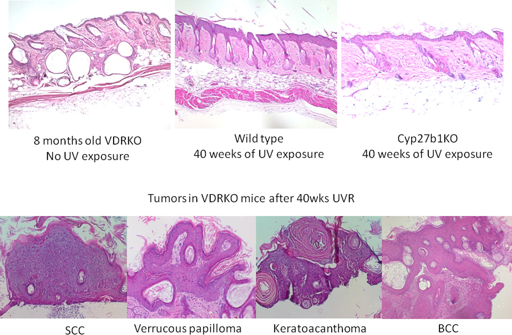
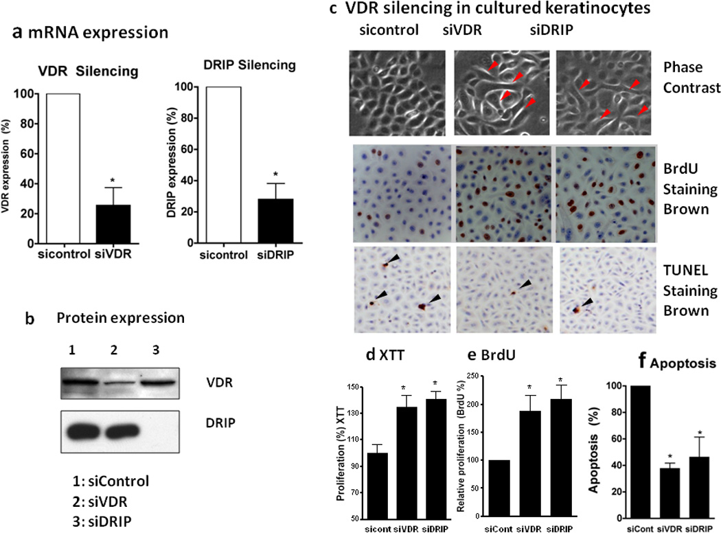
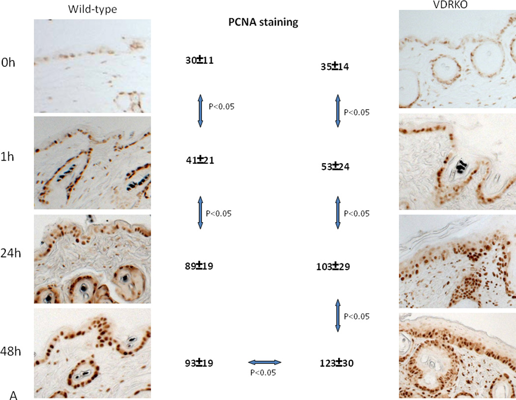
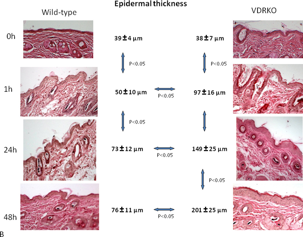
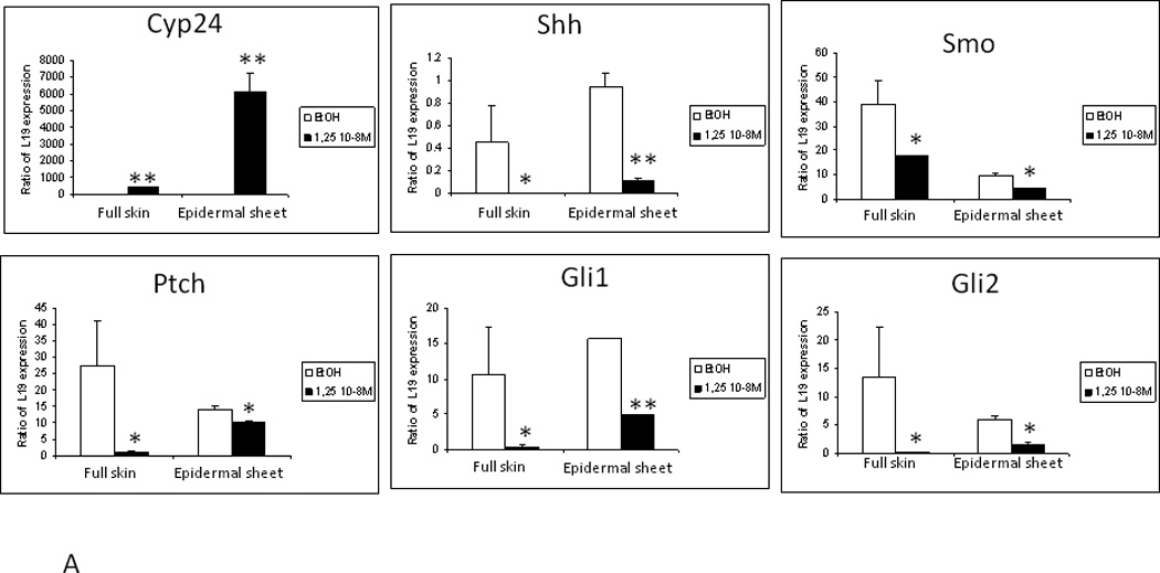
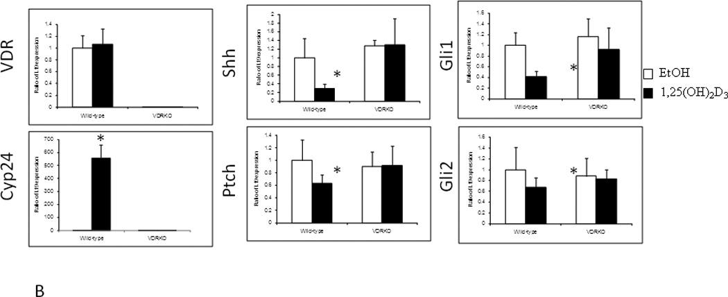
Vitamin D sufficiency is associated with protection against malignancy in a number of tissues clinically, and a strong body of evidence from animal and cell culture studies supports this protective role. Cancers in the skin differ, however, in that higher serum levels of 25OHD are associated with increased basal cell carcinomas (BCC), the most common form of epidermal malignancy. This result may be interpreted as indicating the role of UVR (spectrum 280-320) in producing vitamin D in the skin as well as causing those DNA mutations and proliferative changes that lead to epidermal malignancies. Recent animal studies have shown that mice lacking the vitamin D receptor (VDR) are predisposed to developing skin tumors either from chemical carcinogens such as 7,12-dimethylbenzanthracene (DMBA) or chronic UVR exposure. Such studies suggest that vitamin D production and subsequent signaling through the VDR in the skin may have evolved in part as a protective mechanism against UVR induced epidermal cancer formation. In this manuscript we provide evidence indicating that vitamin D signaling protects the skin from cancer formation by controlling keratinocyte proliferation and differentiation, facilitating DNA repair, and suppressing activation of the hedgehog (Hh) pathway following UVR exposure. This article is part of a Special Issue entitled 'Vitamin D Workshop'.


Figures

Figure 1
Figure 1. Tumors induced by UVR in VDR null mice
A. Representative sections of skin from 8 months old VDR null mice without UVB exposure, wild-type mice after 40 weeks of UVB exposure and CYP27B1 null mice after 40 weeks of UVB exposure. B. Tumors from VDR null mice exposed to 40 weeks of UVB irradiation were collected and classified into papillomas, squamous cell carcinomas (SCC), keratoacanthomas and basal cell carcinomas (BCC). Adapted from Teichert et al. J Invest Dermatol 131:2289-2297, 2011 with permission.
Figure 2
Figure 2. Regulation of keratinocyte proliferation and differentiation by VDR and its coactivators
Epidermal keratinocytes were transfected with non-targeted siRNA for control (sicontrol), VDR (siVDR), and DRIP205 (siDRIP). VDR and DRIP expression was reduced as shown by qRTPCR (a) and western analysis (b). Cells were maintained in low calcium (0.03mM) to keep them proliferative. Cell proliferation was assessed by BrdU incorporation (c, e BrdU) and XTT assay (d XTT). The BrdU incorporated cells (brown) were counted using Bioquant and expressed as % total cells (blue counter staining) (c, e). Keratinocyte apoptosis was evaluated by measuring DNA fragmentation using Apoptaq In situ apoptosis peroxidase detection kit (Chemicon) (c,f TUNEL staining). The brown DNA fragmented cell nuclei (black arrows) per total cells (blue counter staining) were counted. Over 5000 cells were counted in three batches of keratinocytes to make these calculations (f, apoptosis). The hyperproliferation and decreased apoptosis were accompanied by morphologic changes from normal cuboidal epithelial cells tightly aggregated to loosely aggregated spindle shaped cells (red arrows).
Figure 3
Figure 3. Hyperproliferative response to UVR in VDR null epidermis
Wild-type mice exposed to 1 dose of UVB (477 mJ/cm2) showed increased proliferation (A, PCNA staining) and epidermal hyperplasia (B, H&E staining) up to 24h after treatment with no further increase at 48h. VDR null mice exposed to the same dose of UVB showed significantly more pronounced proliferation (A) and epidermal hyperplasia (B) that continued to increase at 48h. Adapted from Teichert et al. J Invest Dermatol 131:2289-2297, 2011 with permission.
Figure 4
Figure 4. Defective DNA Damage Repair in VDR null mouse epidermis following UVR
A. Wildtype and VDR null mice were exposed to 1 dose of UVB (400mJ/cm2) and the skin evaluated for the presence of CPDs over the subsequent 48hrs by immunohistochemistry (anti CPD from Cosmo Biosciences). CPDs were completely cleared by 24hr in the wildtype mouse epidermis, but persisted through 48hrs in the VDR null mouse epidermis. B. The epidermis from 2d old wildtype and VDR null mice was exposed to 35.4mJ/cm2 UVB, and CPDs and 6,4PPs (detected by immunoblots) measured immediately after irradiation and after 46hrs. In the experiment measuring CPDs, half of the epidermal explants were treated with hydroxyurea (HU) to block DNA synthesis prior to and following irradiation. Clearance of CPDs and 6,4PPs was markedly impaired in the VDR null epidermal explants consistent with the in vivo results in A. Adapted from Oh et al. J Invest Dermatol (in press, 2012).
Figure 5
Figure 5. Overexpression of the Hh pathway in VDR null mouse skin and tumors
A. Shh, Ptch1, Smoh, Gli1 and Gli2 proteins as shown by the brown signal were overexpressed in the epidermis and hair follicles of VDR null mice compared to their wild-type littermates at 11 weeks after birth by immunocytochemistry. Slides were counterstained with hematoxylin (blue stain). The bar denotes 50 mm. The protein levels were quantified by western blot. The numerical value represents the average ratio of VDR null band intensity versus wild-type band intensity from three mice per group. * p<0.05. B. Shh, Ptch1, Smoh, Gli1 and Gli2 proteins as detected by immunohistochemistry in a papilloma from a VDR null mouse treated with DMBA and in a BCC from a VDR null mouse treated with UVB. Slides were counterstained with hematoxylin (blue stain). The bar denotes 50 mm. Shh, Ptch1, Smoh, Gli1 and Gli2 protein levels were also measured by western blot in skin tumors and tumor free tissue from DMBA treated VDR null mice. The numerical value represents the mean ratio of the tumor band intensity versus tumor free tissue band intensity from three mice. * p<0.05 Adapted from Teichert et al. J Invest Dermatol 131:2289-2297, 2011 with permission.
Figure 6
Figure 6. Suppression of Hh pathway in mouse skin by 1,25(OH)2D acting through the VDR
A. Treatment of epidermal preparations from wild-type mice in culture with 1,25(OH)2D3 10−8M or EtOH for 24h induced Cyp24 expression and repressed Shh, Gli1, Gli2 and Ptch1 expression. B. Epidermal preparations from wild-type and VDR null mice in culture were treated with 1,25(OH)2D3 10−8M or EtOH for 24h. Absence of VDR expression was verified in VDR null mice, and their epidermis failed to respond to 1,25(OH)2D3 induction of Cyp24 expression unlike that in wild-type mice. 1,25(OH)2D3 treatment repressed Shh, Gli1, Gli2 and Ptch1 expression only in wild-type preparations. * p<0.05. Adapted from Teichert et al. J Invest Dermatol 131:2289-2297, 2011 with permission.

Similar articles

Cited by

References

    1. Eisman JA, Martin TJ, MacIntyre I, et al. 1,25-dihydroxyvitamin-D-receptor in breast cancer cells. Lancet. 1979;2(8156–8157):1335–1336. - PubMed
    1. Ratnam AV, Bikle DD, Su MJ, et al. Squamous carcinoma cell lines fail to respond to 1,25-Dihydroxyvitamin D despite normal levels of the vitamin D receptor. J Invest Dermatol. 1996;106(3):522–525. - PubMed
    1. Kamradt J, Rafi L, Mitschele T, et al. Analysis of the vitamin D system in cutaneous malignancies. Recent Results Cancer Res. 2003;164:259–269. - PubMed
    1. Colston K, Colston MJ, Feldman D. 1,25-dihydroxyvitamin D3 and malignant melanoma: the presence of receptors and inhibition of cell growth in culture. Endocrinology. 1981;108(3):1083–1086. - PubMed
    1. Garland C, Shekelle RB, Barrett-Connor E, et al. Dietary vitamin D and calcium and risk of colorectal cancer: a 19-year prospective study in men. Lancet. 1985;1(8424):307–309. - PubMed

Publication types

MeSH terms