Review
. 2012;88(8):416-33.
doi: 10.2183/pjab.88.416.

Structural studies of large nucleoprotein particles, vaults

Affiliations
Review

Structural studies of large nucleoprotein particles, vaults

Hideaki Tanaka et al. Proc Jpn Acad Ser B Phys Biol Sci. 2012.

Abstract

Vault is the largest nonicosahedral cytosolic nucleoprotein particle ever described. The widespread presence and evolutionary conservation of vaults suggest important biologic roles, although their functions have not been fully elucidated. X-ray structure of vault from rat liver was determined at 3.5 Å resolution. It exhibits an ovoid shape with a size of 40 × 40 × 67 nm(3). The cage structure of vault consists of a dimer of half-vaults, with each half-vault comprising 39 identical major vault protein (MVP) chains. Each MVP monomer folds into 12 domains: nine structural repeat domains, a shoulder domain, a cap-helix domain and a cap-ring domain. Interactions between the 42-turn-long cap-helix domains are key to stabilizing the particle. The other components of vaults, telomerase-associated proteins, poly(ADP-ribose) polymerases and small RNAs, are in location in the vault particle by electron microscopy.


Figures
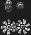
Figure 1.
Figure 1.
Structural models of vault particles based on electron microscopic analyses.10) Top row, Side (left) and top views (right) of intact vault particles. Bottom row, Paired vault flowers derived from a single vault particle. Figures are reproduced from Kedersha et al.10) with copyright permission from the Rockefeller University Press.
Figure 2.
Figure 2.
Cryoelectron microscopic analyses of wild-type vault and vault containing recombinant HisT7-MVP. (A) An isosurface representation of recombinant vault containing HisT7-MVP and (B) a density slice of the image. (C) A density slice of an image of wild-type vault. The two density slices are displayed using the same color map, with stronger densities in red, weaker densities in light blue, and densities at or below background noise in black. Note the weakly dense area in (B) protruding from the inner surface at the central constriction of the particle containing HisT7-MVP. Figures are reproduced from Mikyas et al.2) with copyright permission from Elsevier.
Figure 3.
Figure 3.
Photographs of crystals of vault particles from rat liver. The various panels show different crystal forms, including (A) a crystal (form III), (B) a triclinic crystal (form IV), (C) monoclinic crystals (form V), (D) a monoclinic crystal (form VI), (E) tetragonal crystals (form VII), and (F) crystals (form VIII). Scale bars, 0.1 mm. Figures are reproduced from Kato et al.31) with permission of the International Union of Crystallography.
Figure 4.
Figure 4.
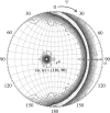
Stereo diagrams based on the slow-rotation function for two-fold rotational symmetry (χ = 180° section) in the 50–10 Å resolution range. Contours were drawn at equal intervals of 0.5σ starting from 0.5σ. The peak at (φ, ψ) = (110°, 90°) represents a multifold rotation axis.
Figure 5.
Figure 5.
R factors (a, R = Σ||Fo| − |Fc||/Σ|Fo|) and correlation coefficients (b, Σ[(〈|Fo|〉 − |Fo|)(〈|Fc|〉 − |Fc|)]/Σ[(〈|Fo|〉 − |Fo|)2(〈|Fc|〉 − |Fc|)2]1/2 plotted against various NCS averaging values at 10 Å resolution. Fo and Fc are observed and calculated structure factors, respectively, and 〈 〉 indicates an averaged value. Figures are reproduced from Kato et al.31) with permission of the International Union of Crystallography.
Figure 6.
Figure 6.
Improvements in electron density during phase extension procedure from 30 Å to 4 Å resolution. Phase extension was performed using NCS averaging based on the 3-fold rotational symmetry. Phase extension was started at 30 Å resolution. The averaged electron density map became clearer as the resolution became higher. The electron density map at 4 Å resolution shows a long helix in the cap domain and repeat structures the body domain.
Figure 7.
Figure 7.
Overall structure of the vault shell based on a ribbon representation of the Cα trace. One MVP molecule is shown in maroon, and the others are shown in green. Left panel, Side view of a vault shell comprising a 78-oligomer polymer of MVP molecules. The particle is ∼670 Å from the top to the bottom, with a maximum diameter of ∼400 Å. The particle has two protruding caps, two shoulders, and a body with an invaginated waist. Two half-vault particles associate at the waist via the N-terminal domains of MVP. Right panel, Top view of the ribbon representation. The maximum diameter of the cap is ∼200 Å. The outer and inner diameters of the cap-ring domain are shown.
Figure 8.
Figure 8.
Left panel, Ribbon representation of an MVP monomer. An MVP monomer contains nine structural repeat domains (R1 to R9), a shoulder domain, a cap-helix domain and a cap-ring domain. N-terminal residue of each domain is represented by numbering along the amino acid sequence. Each domain is depicted in a different color: domain 1 (Met1-Pro55), domain 2 (Arg56-Thr110), domain 3 (Pro111-Ile163), domain 4 (Gln164-Val216), tan; domain 5 (Asp217-Val271), domain 6 (Pro272-Asp322), domain 7 (Val323-Gln378), domain 8 (Ala379-Arg456), domain 9 (Val457-Gly519), shoulder domain (Pro520-Val646), cap-helix domain (Asp647-Leu802) and cap-ring domain (Gly803-Ala845). Middle panels, Two β-sheet topology diagrams of the structural repeat domains. Subgroup A consists of five antiparallel β strands (S1 to S5), whereas subgroup B has additional strands (S2a and S2b) between S2 and S3. The R1 to R7 structure repeat domains belong to subgroup B, whereas R8 and R9 belong to subgroup A. Right panels, Ribbon drawings of R8 (top) and R4 (bottom).
Figure 9.
Figure 9.
A stereoscopic pair of the shoulder domain (Pro520-Val646). From the N-terminal end to the C-terminal end, the secondary structure elements are β1, β2, α1, α2, α3, α4, β3 and β4. A structural model of residues 608–620 has not been built.
Figure 10.
Figure 10.
Ribbon representations of the shoulder domain (left) and SFPH domains from PhStoCD (middle) and FlotBD7 (right). Although the sequence of the shoulder domain is not particularly homologous with that of the SPFH domain from PhStoCD (10.2% identity) or FlotBD7 (6.3% identity), the tertiary structures of the domains are very similar.
Figure 11.
Figure 11.
Location of TEP1 and vRNA. (A) vRNA (red) is located at the ends of the vault caps based on the different density maps for RNase-treated and intact vault reconstructions. (B) Colocalization of TEP1 and vRNA. Vault (red) is shown with the TEP1 WD40 domain (blue) and vRNA (yellow). (C) Modeling of the TEP1 WD40 repeat domain using a portion of the structure of the heterotrimeric G-protein β subunit. (D) A circular β-propeller model of the TEP1 WD40 repeat. Figures are reproduced from Kong et al.9) with copyright permission from the RNA Society.
Figure 12.
Figure 12.
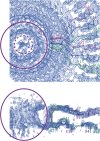
An electron density map of the cap-ring domain (residues 803–845) shown at 2.0 σ. The residue numbers of each molecule are shown in same color as the chain. Top panel, A top view of the map. Bottom panel, A side view of the map. TEP1 is likely reflected in the cylindrical electron density cage enclosed between the purple circle and dashed purple circle in the top panel, and the electron dense area in the purple ellipse in the bottom panel. The cylindrical structure has an outer diameter of ∼45 Å, an inner diameter of ∼20 Å and a height of ∼30 Å. vRNA, which interacts with TEP1,13,14) may be represented by the region enclosed in the red dashed circle in the top panel.
Figure 13.
Figure 13.
VPARP is located in the top half of the vault based on cryoelectron microscopic imaging of recombinant vault particles. (A) A cryoelectron microscopic difference map of VPARP—vault with recombinant HisT7-MVP/VPARP/TEP1 minus vault with recombinant vsvg-MVP/TEP1—is shown in red with the isosurface value set above the level of background noise. (B) The same VPARP difference map shown with the isosurface value set to include weak differences in density and some noise. The three density bands assigned to VPARP are indicated with arrows in (A) and (B). The difference density map is superimposed on the reconstruction of vault with recombinant vsvg-MVP/TEP1 (blue). Figures (A) and (B) are reproduced from Mikyas et al.2) with copyright permission from Elsevier. (C) A vertically sliced section of a ribbon representation with three VPARP sites (arrows). The major site denoted by the lower arrow is inside the R4 structural repeat domain. A minor VPARP site marked with the upper arrow is near Glu717 in the cap-helix domain. A third site denoted by the middle arrow is at the inner surface of the R9 structural repeat domain.
Figure 14.
Figure 14.
Hydrophobic interactions between two adjacent cap-helix domains. The two helices are depicted with ribbon representations and different colors. Amino-acid residues that interact with those from the adjacent helix are identified with single letters and residue numbers.
Figure 15.
Figure 15.
Five ionic interactions between adjacent subunits. Middle panel, Two subunits are shown from the R8 structural repeat domain to the cap-ring domain using ribbon representations. Left panel, Salt bridges between Glu780 and Lys783, and Glu791 and Lys794 in C-terminal regions of the cap-helix domains. Top right panel, A salt bridge between Arg808 and Asp809 in the N-terminal helices of the cap-ring domains. Bottom right panel, Ionic interactions between His675 and Glu681, and Glu689 and Arg703 in N-terminal regions of the cap-helix domains. Amino acids are identified with single letters and residue numbers.
Figure 16.
Figure 16.
Intermolecular interactions between two half-vault particles. N-terminal residues of the R1 structural repeat domain (Met1-Glu4) form an intermolecular antiparallel β sheet with the same residues from the molecule exhibiting two-fold symmetry. Other specific interactions between the two half-vault particles include an ionic bond between Glu4 and Arg42. In contrast to the association between C-terminal cap domains, many N-terminal associations are hydrophilic.
Figure 17.
Figure 17.
A schematic representation of the opening of vault particles. At low pH, acidic residues at the vault interface are neutral, resulting in an electropositive area, reduced ionic attractive force, and disassembly of the vault particle owing to charge repulsion. The half vault moiety on the right of the figure corresponds to the flower-like structures described by Kedersha et al.10) Figures are reproduced from Querol-Audí et al.22) with copyright permission from the Nature Publishing Group.
Figure 18.
Figure 18.
A vertically sliced section of a half vault structure. The capsid structure consisting of MVP molecules is shown in a ribbon representation. Gray circles denote VPARP molecules on the inside of the R4 repeat domain; the molecules are spherical with a diameter of 94 Å. TEP1 in the cap region of vault consists of a 100 × 100 × 80 Å3 circular column and a 45 × 45 × 30 Å3 cylinder (black). The vRNA molecule at the end of the cap region consists of a 15 × 15 × 30 Å3 circular column and an 82 × 82 × 20 Å3 circular disc (gray).

References

    1. Kedersha N.L., Rome L.H. (1986) Isolation and characterization of a novel ribonucleoprotein particle: large structures contain a single species of small RNA. J. Cell Biol. 103, 699–709 - PMC - PubMed
    1. Mikyas Y., Makabi M., Raval-Fernandes S., Harrington L., Kickhoefer V.A., Rome L.H., Stewart P.L. (2004) Cryoelectron microscopy imaging of recombinant and tissue derived vaults: Localization of the MVP N termini and VPARP. J. Mol. Biol. 344, 91–105 - PubMed
    1. Kedersha N.L., Miquel M.C., Bittner D., Rome L.H. (1990) Vaults. II. Ribonucleoprotein structures are highly conserved among higher and lower eukaryotes. J. Cell Biol. 110, 895–901 - PMC - PubMed
    1. Tanaka H., Kato K., Yamashita E., Sumizawa T., Zhou Y., Yao M., Iwasaki K., Yoshimura M., Tsukihara T. (2009) The structure of rat liver vault at 3.5 angstrom resolution. Science 323, 384–388 - PubMed
    1. Kickhoefer V.A., Siva A.C., Kedersha N.L., Inman E.M., Ruland C., Streuli M., Rome L.H. (1999) The 193-kD vault protein, VPARP, is a novel poly(ADP-ribose) polymerase. J. Cell Biol. 146, 917–928 - PMC - PubMed

Publication types

Substances