. 2012 Nov 23;287(48):40652-60.
doi: 10.1074/jbc.M112.419721. Epub 2012 Oct 11.

Voltage-dependent anion channels (VDACs) recruit Parkin to defective mitochondria to promote mitochondrial autophagy

Affiliations

Voltage-dependent anion channels (VDACs) recruit Parkin to defective mitochondria to promote mitochondrial autophagy

Yu Sun et al. J Biol Chem. .

Abstract

Background: Parkin is recruited to defective mitochondria to promote degradation by an autophagy mechanism (mitophagy).

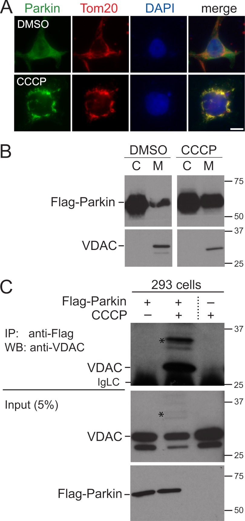
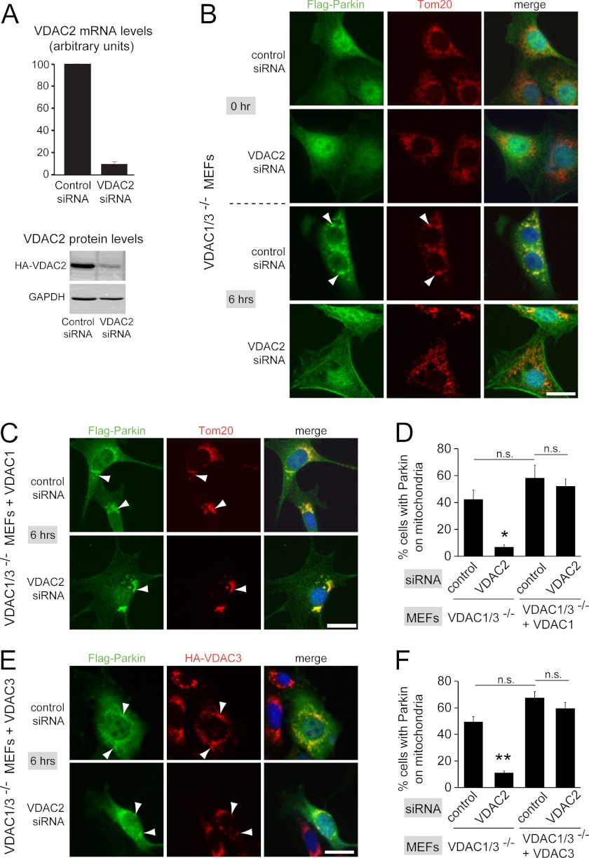
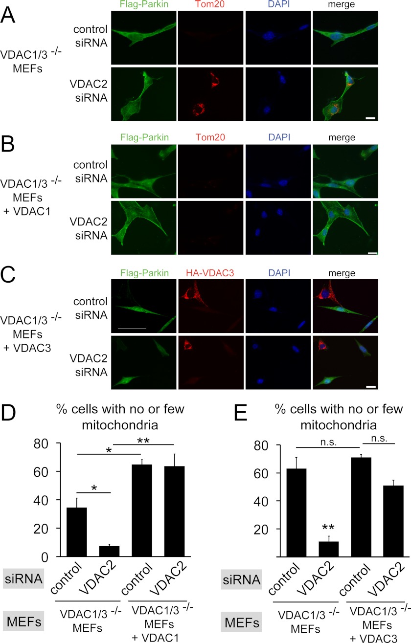
Results: VDACs specifically interact with Parkin on defective mitochondria and are required for efficient targeting of Parkin to mitochondria and subsequent mitophagy.

Conclusion: VDACs recruit Parkin to defective mitochondria.

Significance: A novel mechanistic aspect of Parkin-dependent mitophagy is proposed that may be relevant to Parkinson disease. Mutations in the ubiquitin ligase Parkin and the serine/threonine kinase PINK1 can cause Parkinson disease. Both proteins function in the elimination of defective mitochondria by autophagy. In this process, activation of PINK1 mediates translocation of Parkin from the cytosol to mitochondria by an unknown mechanism. To better understand how Parkin is targeted to defective mitochondria, we purified affinity-tagged Parkin from mitochondria and identified Parkin-associated proteins by mass spectrometry. The three most abundant interacting proteins were the voltage-dependent anion channels 1, 2, and 3 (VDACs 1, 2, and 3), pore-forming proteins in the outer mitochondrial membrane. We demonstrate that Parkin specifically interacts with VDACs when the function of mitochondria is disrupted by treating cells with the proton uncoupler carbonyl cyanide p-chlorophenylhydrazone. In the absence of all three VDACs, the recruitment of Parkin to defective mitochondria and subsequent mitophagy are impaired. Each VDAC is sufficient to support Parkin recruitment and mitophagy, suggesting that VDACs can function redundantly. We hypothesize that VDACs serve as mitochondrial docking sites to recruit Parkin from the cytosol to defective mitochondria.


Figures

FIGURE 1.
FIGURE 1.
Parkin is recruited to mitochondria in response to CCCP treatment and associates with VDAC. A, 293 cells stably expressing FLAG-Parkin were treated for 1 h with 10 μm CCCP or DMSO as a control and immunostained for FLAG-Parkin and the mitochondrial marker Tom20. FLAG-Parkin is normally diffusely localized throughout the cell but is enriched at mitochondria after CCCP treatment. Scale bar, 20 μm. B, as in A except that cells were subjected to subcellular fractionation to generate a cytosol fraction (C) and a membrane fraction enriched in mitochondria (M). FLAG-Parkin and VDACs were detected by Western blotting. Molecular masses are indicated in kDa. C, 293 cells expressing FLAG-Parkin and control 293 cells were treated for 1 h with CCCP or DMSO as a control. Cell lysates were immunoprecipitated (IP) with anti-FLAG antibodies, and associated VDACs were detected with anti-VDAC antibodies by Western blotting (WB). VDACs specifically co-immunoprecipitate with FLAG-Parkin when cells are treated with CCCP. 5% of the lysate used for the immunoprecipitations was loaded for the input. Stars indicate higher molecular mass bands that likely represent covalently modified VDAC. The anti-VDAC antibody was raised against full-length human VDAC1 and cross-reacts with VDAC3 (supplemental Fig. S1B). The minor band below the one marked VDAC may represent VDAC3, which migrates faster than VDAC1 (supplemental Fig. S1B). IgLC indicates the immunoglobulin light chain of the anti-FLAG antibodies.
FIGURE 2.
FIGURE 2.
VDACs are necessary for efficient recruitment of Parkin to defective mitochondria. A, depletion of VDAC2 by siRNA knockdown was confirmed by quantitative PCR (top graph). Measured transcript levels were normalized to β-actin and compared with the nontargeting control siRNA. Bars show the mean of three experiments ± S.E (error bars). To demonstrate VDAC2 depletion at the protein level, VDAC1/3−/− MEFs expressing HA-tagged VDAC2 were treated with siRNAs and analyzed by Western blotting with anti-HA antibodies and anti-GAPDH antibodies as a control. B, VDAC1/3−/− MEFs expressing FLAG-Parkin were transfected with the indicated siRNAs. 48 h after transfection, cells were incubated with 20 μm CCCP for 0 h or 6 h and immunostained for Parkin and the mitochondrial marker Tom20. Punctate FLAG-Parkin structures in the 6 h/control siRNA sample co-localize with mitochondria (arrowheads point to examples). C, as in B, except that VDAC1/3−/− MEFs expressing FLAG-tagged Parkin also expressed HA-VDAC1. Cells were treated with CCCP for 6 h. HA-VDAC1 expression rescues the defective mitochondrial targeting of Parkin caused by depletion of VDAC2. D, the fraction of cells with punctate, mitochondria-localized Parkin after 6 h of CCCP treatment was determined from randomized images of the experiments in B and C. E, as in B, except that VDAC1/3−/− MEFs expressing FLAG-tagged Parkin also expressed HA-VDAC3, and mitochondria were immunostained for HA-VDAC3 instead of Tom20. HA-VDAC3 expression rescues the defective mitochondrial targeting of Parkin caused by depletion of VDAC2. F, quantification as in D, except that images were from experiments shown in E for VDAC1/3−/− MEFs expressing FLAG-tagged Parkin and HA-VDAC3 and from experiments not shown for VDAC1/3−/− MEFs expressing FLAG-tagged Parkin (note that this cell line is different from the one in B and was used to generate VDAC1/3−/− MEFs expressing FLAG-tagged Parkin and HA-VDAC3). Scale bars, 20 μm. Data are means ± S.E. of three independent experiments (>100 cells for each experiment). *, p < 0.02; **, p < 0.001, denoting significant difference from the three other groups. Other comparisons are not significantly different (p > 0.05).
FIGURE 3.
FIGURE 3.
VDAC1 and VDAC3 but not VDAC2 are ubiquitinated in VDAC1/3−/− MEFs. A, VDAC1/3−/− MEFs expressing HA-VDAC1, HA-VDAC2, or HA-VDAC3 were immunostained with anti-HA and anti-Tom20 antibodies. HA-VDACs co-localize with Tom20, indicating correct mitochondrial localization of the HA-tagged VDACs. Scale bar is 10 μm. B, VDAC1/3−/− MEFs expressing FLAG-Parkin and either HA-VDAC1 or HA-VDAC2 were treated for 6 h with 20 μm CCCP or DMSO as a control. Cell lysates were immunoprecipitated (IP) with anti-HA antibodies and analyzed by Western blotting (WB) with anti-ubiquitin antibodies (1st panel) or anti-HA antibodies (2nd panel). Higher molecular mass bands corresponding to ubiquitinated VDACs are detected for HA-VDAC1 when cells are treated with CCCP in the anti-ubiquitin and anti-HA Western blots (Ub-VDAC1). The band marked with an arrowhead in the anti-HA blot is not consistently detected in anti-ubiquitin blots, and the band labeled with an asterisk in the anti-ubiquitin blot is not consistently detected in anti-HA blots. Note that the cell line expressing HA-VDAC2 has somewhat higher expression levels of VDAC and Parkin than the HA-VDAC1 cell line (2nd and 3rd panels). GAPDH was used as a loading control (4th panel). C, VDAC1/3−/− MEFs expressing HA-VDAC1 with or without FLAG-Parkin were treated as in B (lanes 1 and 2). No ubiquitinated, higher molecular mass bands of VDAC1 are detected in the absence of Parkin (lane 1). VDAC1/3−/− MEFs expressing FLAG-Parkin and HA-VDAC3 were treated as in B (lanes 3 and 4). Bands are labeled with an asterisk and arrowhead as in B. Note that HA-VDAC3 migrates faster than HA-VDAC1. IgHC indicates the immunoglobulin heavy chain of the anti-HA antibodies. Molecular masses are indicated in kDa.
FIGURE 4.
FIGURE 4.
VDACs are necessary for efficient elimination of mitochondria. A, VDAC1/3−/− MEFs expressing FLAG-Parkin were transfected with the indicated siRNAs. 48 h after transfection, cells were incubated with 20 μm CCCP for 24 h and immunostained for Parkin and the mitochondrial marker Tom20. Absence of the Tom20 signal indicates that mitochondria are efficiently eliminated in cells transfected with control siRNA, but not when VDAC2 is depleted by siRNA. B, as in A except that HA-VDAC1 was expressed in VDAC1/3−/− MEFs expressing FLAG-tagged Parkin. Expression of HA-VDAC1 rescues the mitochondrial elimination defect caused by VDAC2 depletion. C, as in A except that HA-VDAC3 was expressed in VDAC1/3−/− MEFs expressing FLAG-tagged Parkin and cells were immunostained for HA-VDAC3 instead of Tom20. Expression of HA-VDAC3 rescues the mitochondrial elimination defect caused by VDAC2 depletion. Note that HA-VDAC3 is not expressed in all cells. Scale bars, 20 μm. D, the fraction of cells having no or few detectable mitochondria was determined from randomized images of the experiments in A and B by Tom20 immunofluorescence. Data are means ± S.E. (error bars) of three independent experiments (>100 cells each). *, p < 0.05; **, p < 0.01. E, the fraction of cells having no or few detectable mitochondria was determined from randomized images of the experiments in C and from data not shown for VDAC1/3−/− MEFs expressing FLAG-tagged Parkin. Note that this cell line is different from the one in A and was used to generate VDAC1/3−/− MEFs expressing FLAG-tagged Parkin and HA-VDAC3. This may explain the more efficient mitochondrial elimination compared with D. Data are means ± S.E. of three independent experiments (>100 cells each). **, p < 0.01, significantly different from the three other groups. Other comparisons are not significantly different (p > 0.05).

References

    1. Narendra D. P., Youle R. J. (2011) Targeting mitochondrial dysfunction: role for PINK1 and Parkin in mitochondrial quality control. Antioxid. Redox Signal. 14, 1929–1938 - PMC - PubMed
    1. Springer W., Kahle P. J. (2011) Regulation of PINK1-Parkin-mediated mitophagy. Autophagy 7, 266–278 - PubMed
    1. Lin M. T., Beal M. F. (2006) Mitochondrial dysfunction and oxidative stress in neurodegenerative diseases. Nature 443, 787–795 - PubMed
    1. Schon E. A., Przedborski S. (2011) Mitochondria: the next (neurode) generation. Neuron 70, 1033–1053 - PMC - PubMed
    1. Narendra D., Tanaka A., Suen D. F., Youle R. J. (2008) Parkin is recruited selectively to impaired mitochondria and promotes their autophagy. J. Cell Biol. 183, 795–803 - PMC - PubMed

Publication types

MeSH terms

Substances

LinkOut - more resources