

A case of cystoid macular edema associated with Paclitaxel chemotherapy
- PMID: 23060727
- PMCID: PMC3464324
- DOI: 10.3341/kjo.2012.26.5.388
A case of cystoid macular edema associated with Paclitaxel chemotherapy
Abstract
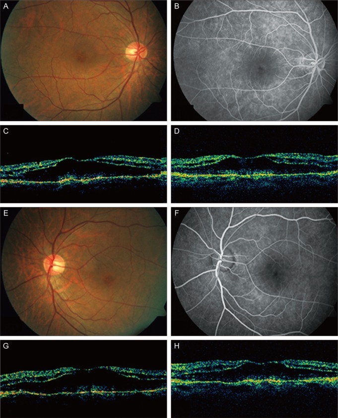
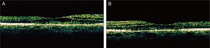
We encountered a patient with cystoid macular edema (CME) secondary to paclitaxel use. A 57-year-old man presented with gradual decreased bilateral vision. His chemotherapeutic regimen consisted of bevacizumab, paclitaxel (175 mg/m(2) for 5 months), and carboplatin. Optical coherence tomography imaging revealed bilateral CME greater than 500 µm. However, one year later, visual acuity was improved, best-corrected Snellen visual acuity was 40 / 80 in each eye, and CME was spontaneously improved. Our study confirmed that macular edema associated with paclitaxel use shows spontaneous resolution and improvement of visual acuity after a change of chemotherapeutic regimen.
Keywords: Macular edema; Paclitaxel.
Conflict of interest statement
No potential conflict of interest relevant to this article was reported.
Figures
References
-
- Tso MO. Pathology of cystoid macular edema. Ophthalmology. 1982;89:902–915. - PubMed
-
- Hofstra LS, de Vries EG, Willemse PH. Ophthalmic toxicity following paclitaxel infusion. Ann Oncol. 1997;8:1053. - PubMed
-
- Teitelbaum BA, Tresley DJ. Cystic maculopathy with normal capillary permeability secondary to docetaxel. Optom Vis Sci. 2003;80:277–279. - PubMed
-
- Telander DG, Sarraf D. Cystoid macular edema with docetaxel chemotherapy and the fluid retention syndrome. Semin Ophthalmol. 2007;22:151–153. - PubMed
-
- Joshi MM, Garretson BR. Paclitaxel maculopathy. Arch Ophthalmol. 2007;125:709–710. - PubMed
Publication types
MeSH terms
Substances
LinkOut - more resources
Full Text Sources
