. 2012 Sep 17:3:157.
doi: 10.3389/fphar.2012.00157. eCollection 2012.

The role of the neuro-astro-vascular unit in the etiology of ataxia telangiectasia

Affiliations

The role of the neuro-astro-vascular unit in the etiology of ataxia telangiectasia

Leenoy Meshulam et al. Front Pharmacol. .

Abstract

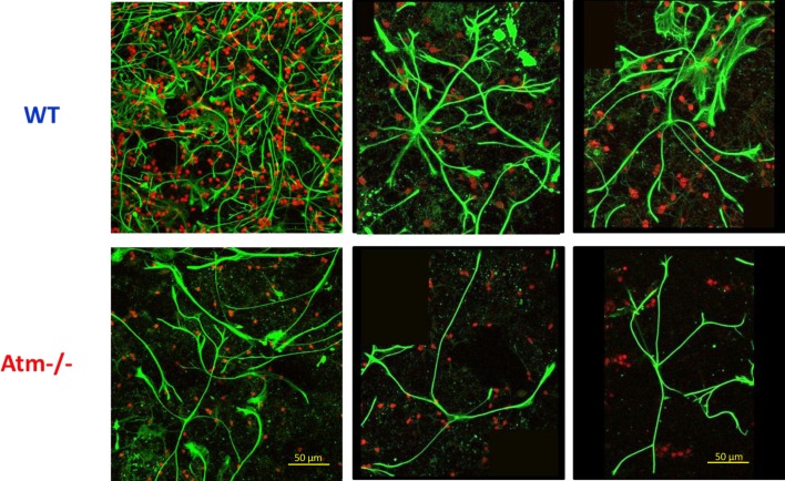
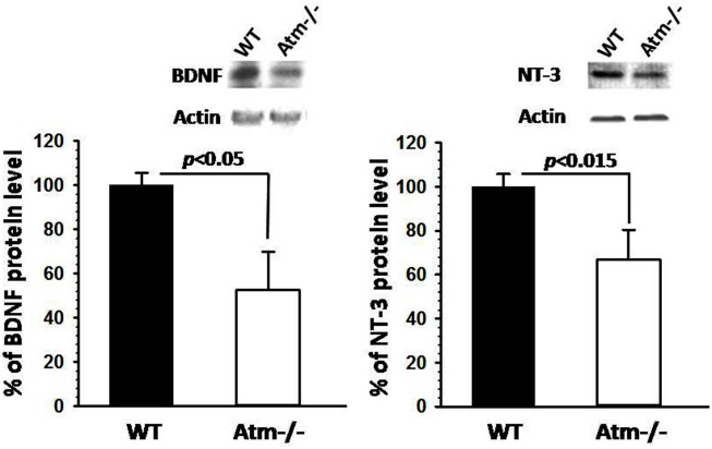
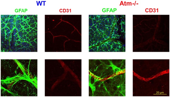
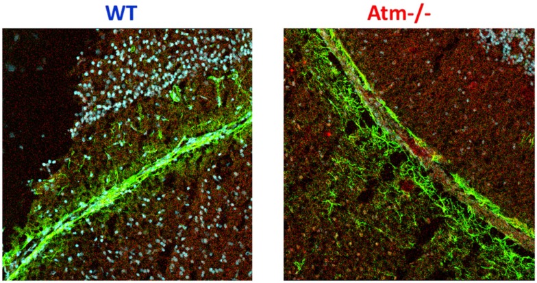
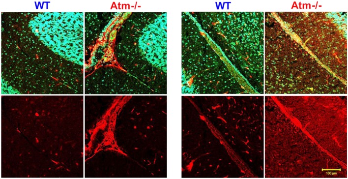
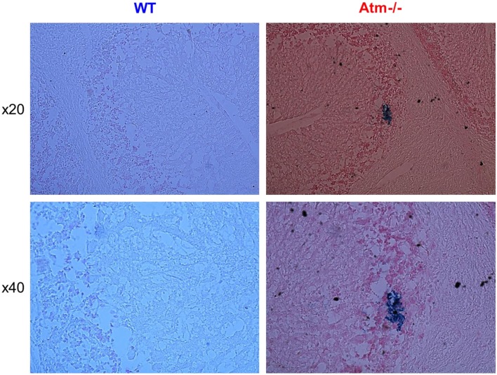
The growing recognition that brain pathologies do not affect neurons only but rather are, to a large extent, pathologies of glial cells as well as of the vasculature opens to new perspectives in our understanding of genetic disorders of the CNS. To validate the role of the neuron-glial-vascular unit in the etiology of genome instability disorders, we report about cell death and morphological aspects of neuroglia networks and the associated vasculature in a mouse model of Ataxia Telangiectasia (A-T), a human genetic disorder that induces severe motor impairment. We found that A-T-mutated protein deficiency was consistent with aberrant astrocytic morphology and alterations of the vasculature, often accompanied by reactive gliosis. Interestingly similar findings could also be reported in the case of other genetic disorders. These observations bolster the notion that astrocyte-specific pathologies, hampered vascularization and astrocyte-endothelium interactions in the CNS could play a crucial role in the etiology of genome instability brain disorders and could underlie neurodegeneration.

Keywords: Ataxia Telangiectasia; DNA damage response; astrocyte; reactive gliosis.


Figures

Figure 1
Figure 1
Glial cell alterations in primary cultures derived from Atm−/− mice. M Immunocytochemical stainingof Glial Fibrillary Acidic Protein (GFAP), a marker of astrocytes (green), and Neuronal Nuclei marker NeuN (red). Note, that Atm−/− astrocytes display significantly less processes in comparison to WT astrocytes. Magnification: left column ×20; center and right column ×40.
Figure 2
Figure 2
Reduction in neurotrophic mediators Atm−/− mice. Western blot analysis displaying reduction in BDNF and NT3 (protein levels in Atm−/− mice (n = 3) and NT3 (n = 3). Error bars represent SEM (statistical analysis was performed using two-tailed Students t test.
Figure 3
Figure 3
Glial cell alterations in retinas of Atm−/− mice. Confocal images of flat-mount retinas from WT and Atm−/− mice, labeled for CD31 (red) and GFAP (green). Magnification: ×4.
Figure 4
Figure 4
Alterations in the glial-vascular interactions in cerebellar sections derived from Atm−/− mice. Confocal images of cerebellar sections from WT and Atm−/− mice, labeled for CD31 (red) and GFAP (green).
Figure 5
Figure 5
Increased fibrinogen expression in retinas of Atm−/− mice. Confocal images of cerebellar sections of mice at 2 months of age show markedly increased fibrinogen immunoreactivity (red) in blood vessels (stained with the pan-endothelial marker CD3, green) of Atm−/− mice compared to the labeling in WT controls. Cell nuclei are stained with Sytox Blue (blue).
Figure 6
Figure 6
Vascular leakage in cerebella of Atm−/− mice. Cerebellar section derived from WT and Atm−/− mice were stained for hemosiderin to indicate deposits resulting from microhemorrhages. The lower panel shows a larger magnification of WT and Atm−/− cerbella. Blue deposits of hemosiderin were evident in cerebella of Atm−/− mice (several indicated with arrows), but scarcely in cerebella of WT mice.

References

    1. Abner C. W., McKinnon P. J. (2004). The DNA double-strand break response in the nervous system. DNA Repair (Amst.) 3, 1141–114710.1016/j.dnarep.2004.03.009 - DOI - PubMed
    1. Andegeko Y., Moyal L., Mittelman L., Tsarfaty I., Shiloh Y., Rotman G. (2001). Nuclear retention of ATM at sites of DNA double strand breaks. J. Biol. Chem. 276, 38224–38230 - PubMed
    1. Antonetti D. A., Barber A. J., Khin S., Lieth E., Tarbell J. M., Gardner T. W. (1998). Vascular permeability in experimental diabetes is associated with reduced endothelial occludin content: vascular endothelial growth factor decreases occludin in retinal endothelial cells. Penn State Retina Research Group. Diabetes 47, 1953–195910.2337/diabetes.47.12.1953 - DOI - PubMed
    1. Arai H., Hori S., Aramori I., Ohkubo H., Nakanishi S. (1990). Cloning and expression of a cDNA encoding an endothelin receptor. Nature 348, 730–73210.1038/348730a0 - DOI - PubMed
    1. Assaf Y., Galron R., Shapira I., Nitzan A., Blumenfeld-Katzir T., Solomon A. S., Holdengreber V., Wang Z. Q., Shiloh Y., Barzilai A. (2008). MRI evidence of white matter damage in a mouse model of Nijmegen breakage syndrome. Exp. Neurol. 209, 181–19110.1016/j.expneurol.2007.09.021 - DOI - PubMed