. 2012 Oct 4:3:307.
doi: 10.3389/fimmu.2012.00307. eCollection 2012.

PAD4 mediated histone hypercitrullination induces heterochromatin decondensation and chromatin unfolding to form neutrophil extracellular trap-like structures

Affiliations

PAD4 mediated histone hypercitrullination induces heterochromatin decondensation and chromatin unfolding to form neutrophil extracellular trap-like structures

Marc Leshner et al. Front Immunol. .

Abstract

NETosis, the process wherein neutrophils release highly decondensed chromatin called neutrophil extracellular traps (NETs), has gained much attention as an alternative means of killing bacteria. In vivo, NETs are induced by bacteria and pro-inflammatory cytokines. We have reported that peptidylarginine deiminase 4 (PAD4), an enzyme that converts Arg or monomethyl-Arg to citrulline in histones, is essential for NET formation. The areas of extensive chromatin decondensation along the NETs were rich in histone citrullination. Here, upon investigating the effect of global citrullination in cultured cells, we discovered that PAD4 overexpression in osteosarcoma U2OS cells induces extensive chromatin decondensation independent of apoptosis. The highly decondensed chromatin is released to the extracellular space and stained strongly by a histone citrulline-specific antibody. The structure of the decondensed chromatin is reminiscent of NETs but is unique in that it occurs without stimulation of cells with pro-inflammatory cytokines and bacteria. Furthermore, histone citrullination during chromatin decondensation can dissociate heterochromatin protein 1 beta (HP1β) thereby offering a new molecular mechanism for understanding how citrullination regulates chromatin function. Taken together, our study suggests that PAD4 mediated citrullination induces chromatin decondensation, implicating its essential role in NET formation under physiological conditions in neutrophils.

Keywords: chromatin decondensation; heterochromatin protein 1; histone modifications; hypercitrullination; neutrophil extracellular traps; pad4.


Figures

Figure 1
Figure 1
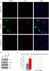
Dramatic chromatin decondensation and formation of NET-like structures upon forced PAD4 expression. (A) Immunostaining of U2OS cells with the H3Cit and the HA antibodies after forced HA-PAD4 expression by transient transfection. Note the dramatic global histone H3 hypercitrullination. (B) Immunostaining of H3Cit and DNA staining showing the enrichment of H3Cit with the highly decondensed chromatin denoted by red arrows. (C) Western blot analyses of the H3Cit levels, and the caspase-3 cleavage in U2OS cells with or without forced HA-PAD4 expression. Histone H3 was probed to ensure equal protein loading.
Figure 2
Figure 2
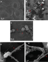
Scanning electron microscope analyses of extracellular chromatin fibers. (A) U2OS cells without forced HA-PAD4 expression. (B–E) U2OS cells with forced HA-PAD4 expression, showing the decondensed chromatin fibers (denoted by red arrows). Also noticeable is the membrane vesicles attached to the chromatin fibers in (E).
Figure 3
Figure 3
PAD4 activity is important for the induction of NET-like structures. (A–C) Fluorescent microscope analyses of histone H3Cit and chromatin morphology in U2OS cells transfected with the pSG5 vector, the pSG5-HA-PAD4 plasmid, or the pSG5-HA-PAD4C645S plasmid. (D) Western blot analyses of HA-fusion protein expression, histone H3Cit levels in U2OS cells after transient transfection. Histone H3 and actin were probed to ensure equal protein loading. (E) The number of H3Cit positive cells without obvious chromatin decondensation (hypercitrullination) or H3Cit positive cells with obvious chromatin decondensation (hypercitrullination and decondensation) were numerated as a percentages of cells that are H3Cit positive in U2OS cells transfected with the pSG5-HA-PAD4 plasmid or the pSG5-HA-PAD4C645S plasmid.
Figure 4
Figure 4
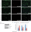
The calcium chelator BAPTA-AM attenuates histone hypercitrullination and chromatin decondensation induced by forced HA-PAD4 expression. (A) Fluorescent microscope analyses of histone H3Cit levels in U2OS cells transfected with the pSG5-HA-PAD4 plasmid and then treated with BAPTA-AM at 0, 5, and 10 μM concentrations. (B) Western blot analyses of histone H3Cit levels in U2OS cells transfected with the pSG5-HA-PAD4 plasmid then treated with BAPTA-AM. Histone H3 blot was performed to show the amount of histone H3 in each sample. HA western blot was performed to monitor the HA-PAD4 expression. (C) The number of H3Cit positive cells without obvious chromatin decondensation (citrullination only) or H3Cit positive cells with obvious chromatin decondensation (citrullination and decondensation) were numerated as a percentages of cells that are H3Cit positive in U2OS cells transfected with the pSG5-HA-PAD4 plasmid and then treated with BAPTA-AM at different concentrations.
Figure 5
Figure 5
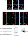
Forced HA-PAD4 expression induced heterochromatin decondensation and HP1β dissociation in NIH 3T3 cells. (A) Immunostaining assays of H3Cit and HP1β in NIH 3T3 cells after forced HA-PAD4 expression. Arrows denote a cell with an increase in H3Cit, a loss of HP1β and the organization of distinct heterochromatic loci, a distinct feature of these cells. (B) Immunostaining with H3Cit and HP1 β in NIH 3T3 cells with forced HA-PAD4 expression. Note the extreme chromatin decondensation in the extracellular space. (C) The sequence of the biotin conjugated H3 N-terminal peptides with no modification, K9me3, or Cit8K9me3 modifications. (D) Coomassie blue staining to show the amount of each peptide. (E) Peptide pull-down experiments to analyze the binding of HP1β to H3 unmod, K9me3, and Cit8K9me3 peptides. Top panel shows a short exposure and bottom panel shows a long exposure time in Western blot assays.
Figure 6
Figure 6
Working model for the effects of H3Arg8 (R8) citrullination on the binding of HP1β to H3K9me3. (1) Upon methylation of H3K9 by the methyltransferase (e.g., Suv39), HP1β recognizes this modification and is recruited to a chromatin region to regulate heterochromatin structure and gene repression. (2) Reversely, PAD4-catalyzed citrullination of H3R8 produce a dual histone H3 modification—H3Cit8K9me3—to dissociate HP1 and mediate heterochromatin decondensation during NET formation.

References

    1. Bianchi M., Hakkim A., Brinkmann V., Siler U., Seger R. A., Zychlinsky A., et al. (2009). Restoration of NET formation by gene therapy in CGD controls aspergillosis. Blood 114, 2619–2622 10.1182/blood-2009-05-221606 - DOI - PMC - PubMed
    1. Bock I., Kudithipudi S., Tamas R., Kungulovski G., Dhayalan A., Jeltsch A. (2011). Application of Celluspots peptide arrays for the analysis of the binding specificity of epigenetic reading domains to modified histone tails. BMC Biochem. 12, 48 10.1186/1471-2091-12-48 - DOI - PMC - PubMed
    1. Brinkmann V., Reichard U., Goosmann C., Fauler B., Uhlemann Y., Weiss D. S., et al. (2004). Neutrophil extracellular traps kill bacteria. Science 303, 1532–1535 10.1007/978-1-61779-527-5_7 - DOI - PubMed
    1. Eissenberg J. C., Elgin S. C. R. (2000). The HP1 protein family: getting a grip on chromatin. Curr. Opin. Genet. Dev. 10, 204–210 10.1016/S0959-437X(00)00058-7 - DOI - PubMed
    1. Fischle W., Tseng B. S., Dormann H. L., Ueberheide B. M., Garcia B. A., Shabanowitz J., et al. (2005). Regulation of HP1-chromatin binding by histone H3 methylation and phosphorylation. Nature 438, 1116–1122 10.1038/nature04219 - DOI - PubMed