

Microleakage comparison of four dental materials as intra-orifice barriers in endodontically treated teeth
- PMID: 23060910
- PMCID: PMC3467124
Microleakage comparison of four dental materials as intra-orifice barriers in endodontically treated teeth
Abstract
Introduction: The aim of this in vitro study was to compare polymicrobial microleakage of calcium enriched mixture (CEM) cement, mineral trioxide aggregate (MTA), amalgam, and composite resin as intra-orifice sealing materials.
Materials and methods: Seventy single-rooted mandibular premolars were instrumented and obturated by cold lateral compaction technique. The teeth were randomly divided into four experimental groups according to used material: CEM, MTA, amalgam and composite resin (n=15) and two control groups (n=5). In experimental groups, 2 mm of coronal gutta-percha was removed and replaced with the study material. All the teeth were mounted in a two-chamber apparatus and the coronal portion was exposed to human saliva. The day the turbidity occurred was recorded for each sample. Data were analyzed using one-way ANOVA.
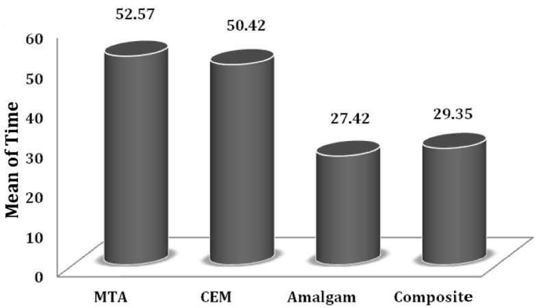
Results: The negative control group showed no leakage while the average microleakage time in the positive control group was 3.5 days. The average bacterial leakage times for amalgam, composite resin, MTA, and CEM groups were 27.42±3.6, 29.35±3.15, 52.57±2.87, and 50.42±2.73 days, respectively. There was no significant difference between CEM and MTA groups (P=0.27) and also between amalgam and composite resin groups (P=0.36). However, in term of average leakage time, MTA and CEM groups exhibited significant differences with amalgam and composite resin groups (P<0.001).
Conclusion: According to the results of the present in vitro study, in terms of coronal sealing in endodontically treated teeth, CEM and MTA are more effective than amalgam and composite resin.
Keywords: Amalgam; Calcium Enriched Mixture; Dental Leakage; Endodontics; MTA; Saliva.
Conflict of interest statement
Figures
Similar articles
-
In Vitro Microleakage of Mineral Trioxide Aggregate, Calcium-Enriched Mixture Cement and Biodentine Intra-Orifice Barriers.Iran Endod J. 2017 Spring;12(2):211-215. doi: 10.22037/iej.2017.41. Iran Endod J. 2017. PMID: 28512488 Free PMC article.
-
Apical Microleakage in Root Canals Containing Broken Rotary Instruments.Iran Endod J. 2017 Summer;12(3):360-365. doi: 10.22037/iej.v12i3.16656. Iran Endod J. 2017. PMID: 28808466 Free PMC article.
-
Evaluating the Effect of Resection on the Sealing Ability of MTA and CEM Cement.Iran Endod J. 2012 Summer;7(3):134-8. Epub 2012 Aug 1. Iran Endod J. 2012. PMID: 23056132 Free PMC article.
-
Orifice barriers to prevent coronal microleakage after root canal treatment: systematic review and meta-analysis.Aust Dent J. 2023 Jun;68(2):78-91. doi: 10.1111/adj.12951. Epub 2023 Feb 9. Aust Dent J. 2023. PMID: 36661351
-
Evaluation of coronal microleakage of intra-orifice barrier materials in endodontically treated teeth: A systematic review.J Conserv Dent. 2022 Nov-Dec;25(6):588-595. doi: 10.4103/jcd.jcd_377_22. Epub 2022 Oct 13. J Conserv Dent. 2022. PMID: 36591578 Free PMC article. Review.
Cited by
-
The Effect of Different Mixing Methods on the Properties of Calcium-enriched Mixture Cement: A Systematic Review of in Vitro Studies.Iran Endod J. 2019 Fall;14(4):240-246. doi: 10.22037/iej.v14i4.25126. Iran Endod J. 2019. PMID: 36794105 Free PMC article. Review.
-
An In-Vitro Analysis of Root Fracture Strength Using Single File Systems.Cureus. 2023 Feb 26;15(2):e35477. doi: 10.7759/cureus.35477. eCollection 2023 Feb. Cureus. 2023. PMID: 36999110 Free PMC article.
-
Double Antibiotic Paste for Management of External Inflammatory Root Resorption.Iran Endod J. 2018 Fall;13(4):569-572. doi: 10.22037/iej.v13i4.22893. Iran Endod J. 2018. PMID: 36883027 Free PMC article.
-
Effect of MTA and CEM on Mineralization-Associated Gene Expression in Stem Cells Derived from Apical Papilla.Iran Endod J. 2018 Winter;13(1):94-101. doi: 10.22037/iej.v13i1.17860. Iran Endod J. 2018. PMID: 29692843 Free PMC article.
-
Importance and methodologies of endodontic microleakage studies: A systematic review.J Clin Exp Dent. 2017 Jun 1;9(6):e812-e819. doi: 10.4317/jced.53604. eCollection 2017 Jun. J Clin Exp Dent. 2017. PMID: 28638561 Free PMC article. Review.
References
-
- Tselnik M, Baumgartner JC, Marshall JG. Bacterial leakage with mineral trioxide aggregate or a resin-modified glass ionomer used as a coronal barrier. J Endod. 2004;30(11):782–4. - PubMed
-
- Roghanizad N, Jones JJ. Evaluation of coronal microleakage after endodontic treatment. J Endod. 1996;22(9):471–3. - PubMed
-
- Chailertvanitkul P, Saunders W, Saunders E, MacKenzie D. An evaluation of microbial coronal leakage in the restored pulp chamber of root canal treated multirooted teeth. Int Endod J. 1997;30(5):318–22. - PubMed
-
- Beckham BM, Anderson RW, Morris CF. An evaluation of three materials as barriers to coronal microleakage in endodontically treated teeth. J Endod. 1993;19(8):388–91. - PubMed
-
- Luketic SF, Malcic A, Jukic S, Anic I, Segovic S, Kalenic S. Coronal microleakage of two root-end filling materials using a polymicrobial marker. J End. 2008;34(2):201–3. - PubMed
LinkOut - more resources
Full Text Sources