. 2012:3:98.
doi: 10.4103/2152-7806.100187. Epub 2012 Aug 27.

"Time is brain" the Gifford factor - or: Why do some civilian gunshot wounds to the head do unexpectedly well? A case series with outcomes analysis and a management guide

Affiliations

"Time is brain" the Gifford factor - or: Why do some civilian gunshot wounds to the head do unexpectedly well? A case series with outcomes analysis and a management guide

David J Lin et al. Surg Neurol Int. 2012.

Abstract

Background: Review of intracranial gunshot wounds (GSWs) undergoing emergent neurosurgical intervention despite a very low Glasgow Coma Scale (GCS) score on admission in order to identify predictors of good outcome, with correlates to recent literature.

Methods: A retrospective review of select cases of GSWs presenting to our trauma center over the past 5 years with poor GCS requiring emergent neurosurgical intervention and a minimum of 1-year follow-up.

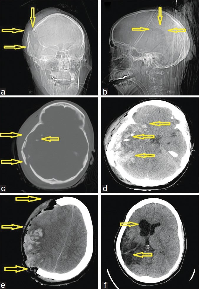
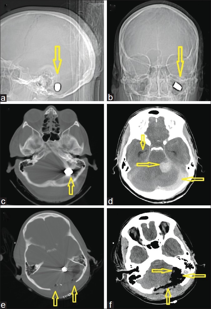
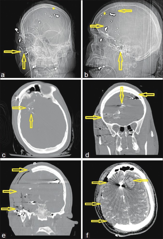
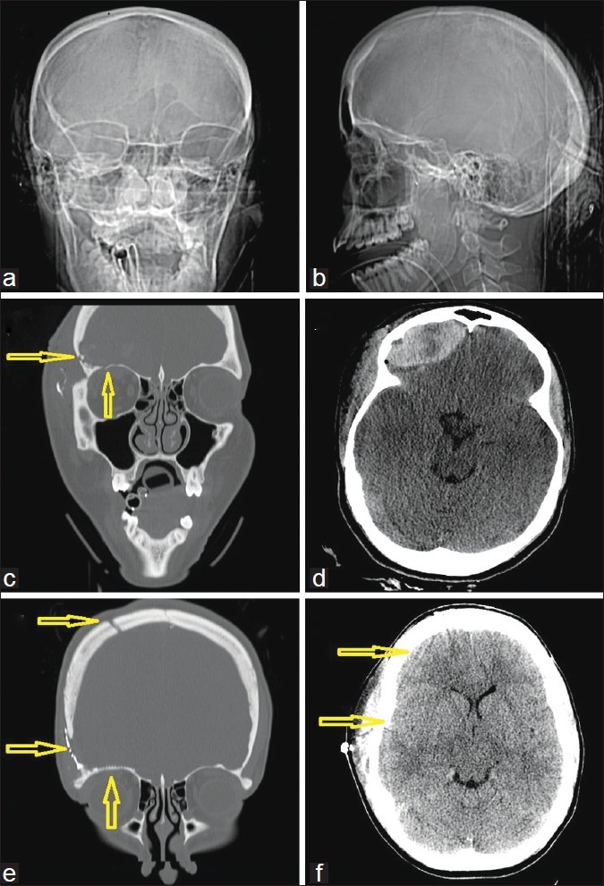
Results: Out of a total of 17 patients who went to the operating room (OR) for GSW to the head during this period, 4 cases with a GCS < 5 on admission were identified. All cases required a hemicraniectomy to alleviate cerebral swelling. Two cases presented with a unilaterally blown pupil due to raised intracranial pressure. The remaining 2 cases had equal and reactive pupils. One patient with a GCS of 3 and a significant bilateral pattern of parenchymal bullet injury was initially assessed in moribund status but rallied and received a delayed hemicraniectomy on day 7. Three out of 4 patients are functionally independent at 1-year follow-up. The fourth patient who received a delayed decompression remains wheelchair dependent.

Conclusion: Victims of GSWs can have good outcomes despite having a very poor admission GCS score and papillary abnormalities. Factors predicting good outcomes include the following: time from injury to surgical intervention of < 1 h; injury to noneloquent brain; and absence of injury to midbrain, brainstem, and major vessels.

Keywords: Decompressive hemicraniectomy; gunshot wounds; head; intracranial.


Figures

Figure 1
Figure 1
A 25-year-old woman with a gunshot wound to the right cerebral hemisphere. (a) computed tomography (CT)-scout image anterior view and (b) lateral view. Arrows demonstrate the right parietal blowout fracture representing the exit wound. (c) Admission CT, axial views in bone windows, clearly showing the right side shattered calvaria, which was likely acting as a hinge-craniotomy thus accommodating some swelling and at the same level (d) soft tissue windows, demonstrating the extensive multilobar hemorrhagic contusions caused by the pressure wave. (e) Immediate postoperative scan after a wide hemicraniectomy with expansile onlay duroplasty. (f) Postoperative scan after the patient had undergone delayed reconstruction with an allograft cranioplasty as indicated by the arrows
Figure 2
Figure 2
A 30-year-old woman with a gunshot wound to the back of the head. (a) computed tomography (CT)-scout image anterior view and (b) lateral view. Arrows demonstrate the left suboccipital target site with an intact bullet (c) Admission CT, axial views in bone windows, showing the bullet which was lodged behind the petrous bone (d) Soft tissue windows, demonstrating the focal posterior fossa hemorrhage near the cerebellopontine angle, but leaving an intact brain stem. A second significant hemorrhage/collection under the occipital bone is visible possibly originating from the sinus. Note the dilated temporal horns bilaterally indicative of obstructive hydrocephalus prior to external ventricular drain insertion. (e) Immediate postoperative scan after a wide bilateral suboccipital midline craniectomy with expansile onlay duroplasty. (f) This scan shows a postoperative scan of the same slice location in soft tissue windows after decompression as indicated by the arrows
Figure 3
Figure 3
A 26-year-old man with a gunshot wound to the right ear and orbit. (a) Computed tomography (CT)-scout image anterior view and (b) lateral view. Double arrows demonstrate the entry site at the right zygoma with a completely disintegrated bullet. (c) Admission CT, axial views, bone windows, showing the metal artifacts from the bullet case, which was scattered along the sphenoid bone. (d) Preoperative coronal reconstructions of the bone windows, demonstrating the midline crossing bullet trajectory as indicated by the arrows (star represents ricochet point). Note that the path does not cross a sinus or the ventricles. (e) Immediate postoperative result also in coronal reconstructions after a wide right hemicraniectomy with duroplasty. (f) Postoperative CT scan on the same day in soft tissue windows after decompression as indicated by the arrows. Note the new left frontal hemorrhagic contusion from the bullet fragment that was bounced off the inner table at the ricochet point
Figure 4
Figure 4
A 26-year-old woman with a penetrating gunshot wound to the head. (a) Computed tomography (CT)-scout image anterior view and (b) lateral view. Several arrows demonstrate the right frontoparietal entry site and a completely disintegrated bullet. (c) Admission CT, axial views, bone windows, showing the metal artifacts from the bullet case, which was scattered along the path. (d) Admission CT, axial views, corresponding soft tissue windows, also showing metal artifacts from the bullet case as well as bone fragments, scattered along the path and some perifocal hypodensity likely indicating edema. (e) Preoperative CT, coronal reconstructions in bone windows, demonstrating the midline crossing bullet trajectory from left to right as indicated by the arrows. Note that the matching CT venogram in (f) shows that the path did not cross the superior sinus or the ventricle

References

    1. Aldrich EF, Eisenberg HM, Saydjari C, Foulkes MA, Jane JA, Marshall LF, et al. Predictors of mortality in severely head-injured patients with civilian gunshot wounds: A report from the NIH Traumatic Coma Data Bank. Surg Neurol. 1992;38:418–23. - PubMed
    1. Benzel EC, Day WT, Kesterson L, Willis BK, Kessler CW, Modling D, et al. Civilian craniocerebral gunshot wounds. Neurosurgery. 1991;29:67–71. discussion 71-2. - PubMed
    1. Berlit P, Jaschke W, Tornow K. [Gunshot injuries of the skull.Computerized tomography findings and clinical course] Nervenarzt. 1987;58:300–4. - PubMed
    1. (CDC) CfDCaP. Nonfatal and fatal firearm-related injuries - United States. MMWR Morb Mortal Wkly Rep. 1999;48:1029–34. - PubMed
    1. Clark WC, Muhlbauer MS, Watridge CB, Ray MW. Analysis of 76 civilian craniocerebral gunshot wounds. J Neurosurg. 1986;65:9–14. - PubMed