. 2012 Jun;2(2):87-97.
doi: 10.3978/j.issn.2223-4292.2012.06.06.

Ultrasound for molecular imaging and therapy in cancer

Affiliations

Ultrasound for molecular imaging and therapy in cancer

Osamu F Kaneko et al. Quant Imaging Med Surg. 2012 Jun.

Abstract

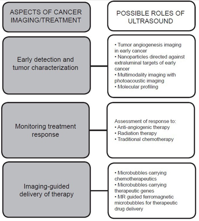
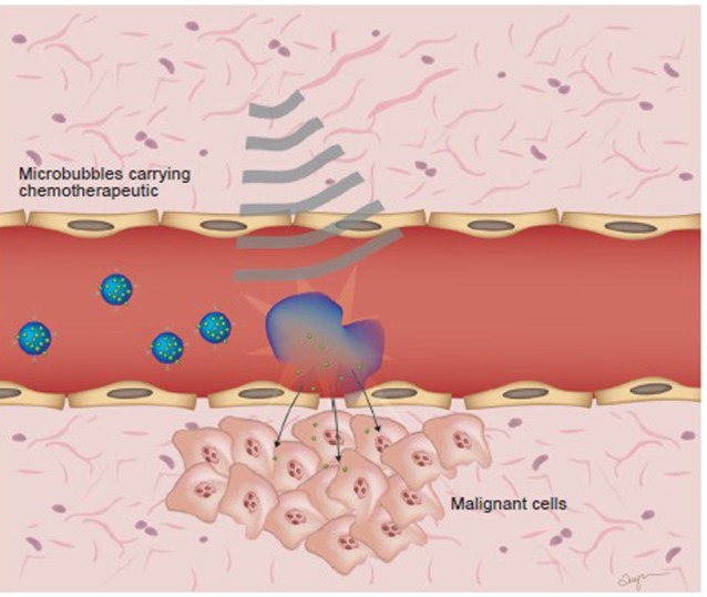
Over the past decade, molecularly-targeted contrast enhanced ultrasound (ultrasound molecular imaging) has attracted significant attention in preclinical research of cancer diagnostic and therapy. Potential applications for ultrasound molecular imaging run the gamut from early detection and characterization of malignancies to monitoring treatment responses and guiding therapies. There may also be a role for ultrasound contrast agents for improved delivery of chemotherapeutic drugs and gene therapies across biological barriers. Currently, a first Phase 0 clinical trial in patients with prostate cancer assesses toxicity and feasibility of ultrasound molecular imaging using contrast agents targeted at the angiogenic marker vascular endothelial growth factor receptor type 2 (VEGFR2). This mini-review highlights recent advances and potential applications of ultrasound molecular imaging and ultrasound-guided therapy in cancer.


Figures

Figure 1
Figure 1
Potential applications of ultrasound molecular imaging in relation to different aspects of imaging. Ultrasound molecular imaging could potentially be used in most aspects of cancer imaging, spanning from early detection to monitoring treatment effects and also for the delivery of chemotherapeutics and gene therapies. Different potential applications are listed adjacent to different phases of cancer imaging
Figure 2
Figure 2
Different ultrasound contrast agents and their distribution relative to the vascular endothelial cells. A: Schematic representation of different micro- and nanoparticles and their size ranges. Microbubbles are typically composed of a phospholipid shell encircling a gas, such as perfluorocarbon (PFC) and total size ranges from 1-4 µm. Targeting of microbubbles is achieved by incorporating for example peptides or antibodies into the microbubble shell. Smaller agents include PFC nanodroplets, which are similar in composition to microbubbles but range from 200-400 nm in size. Polylactic acid has also been used for the shell to make small nanoparticle agents. Solid nanoparticles range between 20-100 nm and may be detectable by ultrasound due to the small amounts of gas trapped within cavities. Liposomes, ranging from 20 nm -10 µm consist of an amphiphilic bilayer surrounding an aqueous core. B: Microbubbles due to their size are restricted to the intravascular space. Targeted microbubbles interact with target ligands on the endothelium. Smaller nanoparticle contrast agents are able to extravasate through the endothelium and enter the extravascular space, which opens up possibilities for targeting extraluminal molecules
Figure 3
Figure 3
Nanobubbles coalescing in the extravascular space become detectable by ultrasound. Nanobubbles due to their small size are able to extravasate through the vascular endothelium, possibly in part due to defective tumor neovasculature. It has been proposed that once in the extravascular space, nanobubbles can coalesce into micron sized collections, thus allowing for ultrasound detectability
Figure 4
Figure 4
Ultrasonic molecular imaging of tumor angiogenesis in breast cancer. Transverse ultrasound image of orthotopically implanted human breast adenocarcinoma xenograft (MDA-MB-231 cells) in nude mouse imaged following intravenous administration of 5×10⁷ VEGFR2-targeted contrast microbubbles (BR55). Note strong imaging signal in breast cancer compared to surrounding normal tissue due to increased angiogenesis and up-regulation of VEGFR2 on angiogenic vascular endothelial cells in breast cancer. Scale bar =3 mm
Figure 5
Figure 5
Schematic representation of microbubble contrast agents as therapeutic drug vehicles. Microbubbles carrying chemotherapeutic drugs or genetic payloads for gene therapies are imaged and bursted at the site of the tumor, resulting in sonoporation and delivery of the drug directly to the tumor site. The process of sonoporation transiently increases cell membrane permeability which facilitates the uptake of extracellular macromolecule chemotherapeutics and, in the case of gene therapy, gene-encoding plasmids

References

    1. Wilson SR, Burns PN. Microbubble-enhanced US in body imaging: what role? Radiology 2010;257:24-39 - PubMed
    1. Pysz MA, Willmann JK. Targeted contrast-enhanced ultrasound: an emerging technology in abdominal and pelvic imaging. Gastroenterology 2011;140(3):785-90 - PMC - PubMed
    1. Piscaglia F, Bolondi L.The safety of Sonovue in abdominal applications: retrospective analysis of 23188 investigations. Ultrasound Med Biol 2006;32:1369-75 - PubMed
    1. Deshpande N, Needles A, Willmann JK. Molecular ultrasound imaging: current status and future directions. Clin Radiol 2010;65:567-81 - PMC - PubMed
    1. Kircher MF, Willmann JK. Molecular Body Imaging: MR Imaging, CT, and US. Part I. Principles. Radiology 2012;263:633-43 - PMC - PubMed