Review
. 2012 Sep;32(5):463-70.
doi: 10.1016/j.semnephrol.2012.07.009.

Targeting pericyte differentiation as a strategy to modulate kidney fibrosis in diabetic nephropathy

Affiliations
Review

Targeting pericyte differentiation as a strategy to modulate kidney fibrosis in diabetic nephropathy

Benjamin D Humphreys. Semin Nephrol. 2012 Sep.

Abstract

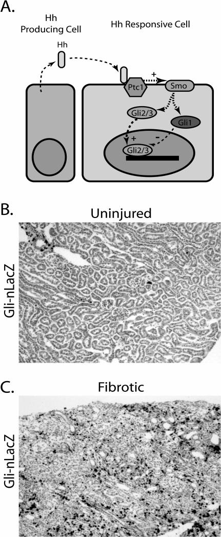
Pericytes are a heterogeneous group of extensively branched cells located in microvessels where they make focal contacts with endothelium. Pericytes stabilize blood vessels, regulate vascular tone, synthesize matrix, participate in repair, and serve as progenitor cells, among other functions. Recent work has highlighted the role of pericytes and pericyte-like cells in fibrosis, in which chronic injury triggers pericyte proliferation and differentiation into collagen-secretory, contractile myofibroblasts with migration away from vessels, causing microvascular rarefaction. In this review the developmental origins of kidney pericytes and perivascular fibroblasts are summarized, pericyte to myofibroblast transition in type I diabetic nephropathy is discussed, and the regulation of pericyte differentiation into myofibroblasts as a therapeutic target for treatment of diabetic nephropathy is described.


Figures

Figure 1
Figure 1. Kidney Pericyte Morphology
A. Electron micrograph of pericyte processes with pale cytoplasm (#) encircling endothelial cells in tumor stroma (*). Note that these pericyte processes lie underneath the capillary basement membrane. Image courtesy of Brian Eyden, PhD, reprinted with permission.B. Fluorescence image of adult mouse kidney interstitium from a FoxD1-GFPCre; R26tdTomato bigenic mouse. Note the finely branched processes emanating from the pericyte cell bodies, extending around tubular basement membrane. C. An interstitial kidney pericyte encircling a peritubular capillary, with the endothelial cell nucleus visible (*).
Figure 2
Figure 2. Myofibroblast Morphology
Electron micrograph of a myofibroblast, with stellate appearance. This cell has abundant myofilaments in cytoplasm (*) as well as dense fibronectin along the cell surface (arrows). Image courtesy of Brian Eyden, PhD, reprinted with permission.
Figure 3
Figure 3. Pericyte to Myofibroblast Transition
A. Genetically labeled pericytes with fine process identified in fluorescence in a FoxD1-GFPCre; R26tdTomato bigenic mouse kidney. B. Ten days after unilateral ureteral obstruction, there is an expansion of labeled cells in interstitium, reflecting proliferation and differentiation of pericytes into myofibroblasts during chronic injury.
Figure 4
Figure 4. Myofibroblast Origins and Signaling
Cartoon depicting the developmental origins of pericytes and fibroblasts in kidney from a FoxD1+ progenitor in mesenchyme during development. In the adult, pericytes can be activated by various growth factors to differentiate into myofibroblasts. Whether adult pericytes (or a subset) have the potential to differentiate into other cell types besides myofibroblasts, as do some pericytes in other vascular beds, remains an open question in kidney.
Figure 5
Figure 5. Hedgehog-Gli1 Signaling in Fibrosis
A. Hh ligand is produced by one cell and acts in a paracrine fashion by binding to Ptch1 on a different cell. This binding releases tonic inhibition of Smo by Ptch1. Smo subsequently activates Gli1-3 effectors, and Gli2/3 translocates to the nucleus where it activates target gene transcription. B. Kidney cortex from a Gli1-nLacZ reporter mouse at ten weeks, stained with Xgal. C. After unilateral ureteral obstruction there is a dramatic upregulation of Gli1-nLacZ expression in kidney interstitium, reflecting activation of the Hh-Gli pathway in kidney pericytes (Fabian and Humphreys, unpublished).

References

    1. Risdon RA, Sloper JC, De Wardener HE. Relationship between renal function and histological changes found in renal-biopsy specimens from patients with persistent glomerular nephritis. Lancet. 1968;2:363–366. - PubMed
    1. Nath KA. Tubulointerstitial changes as a major determinant in the progression of renal damage. Am J Kidney Dis. 1992;20:1–17. - PubMed
    1. Katz A, Caramori ML, Sisson-Ross S, et al. An increase in the cell component of the cortical interstitium antedates interstitial fibrosis in type 1 diabetic patients. Kidney Int. 2002;61:2058–2066. - PubMed
    1. Fioretto P, Mauer M, Brocco E, et al. Patterns of renal injury in NIDDM patients with microalbuminuria. Diabetologia. 1996;39:1569–1576. - PubMed
    1. Essawy M, Soylemezoglu O, Muchaneta-Kubara EC, et al. Myofibroblasts and the progression of diabetic nephropathy. Nephrol Dial Transplant. 1997;12:43–50. - PubMed

Publication types

MeSH terms