. 2012 Oct 12;151(2):253-66.
doi: 10.1016/j.cell.2012.09.024.

Pathogenic simian immunodeficiency virus infection is associated with expansion of the enteric virome

Affiliations

Pathogenic simian immunodeficiency virus infection is associated with expansion of the enteric virome

Scott A Handley et al. Cell. .

Abstract

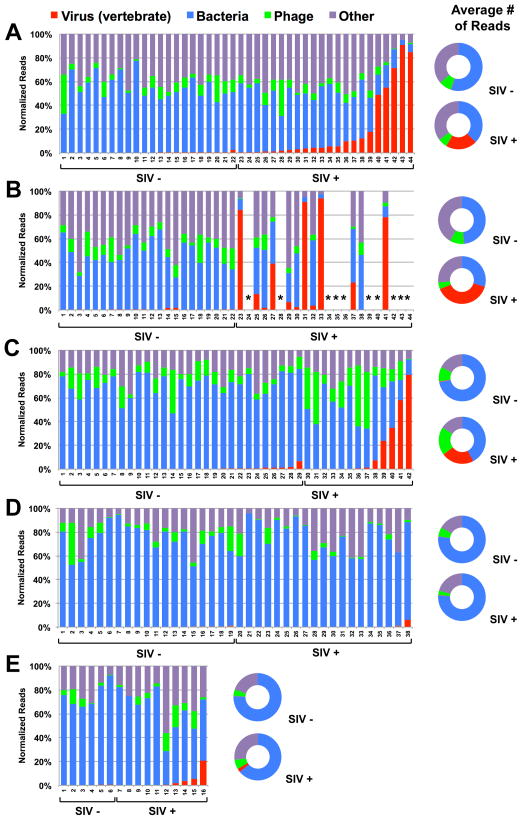
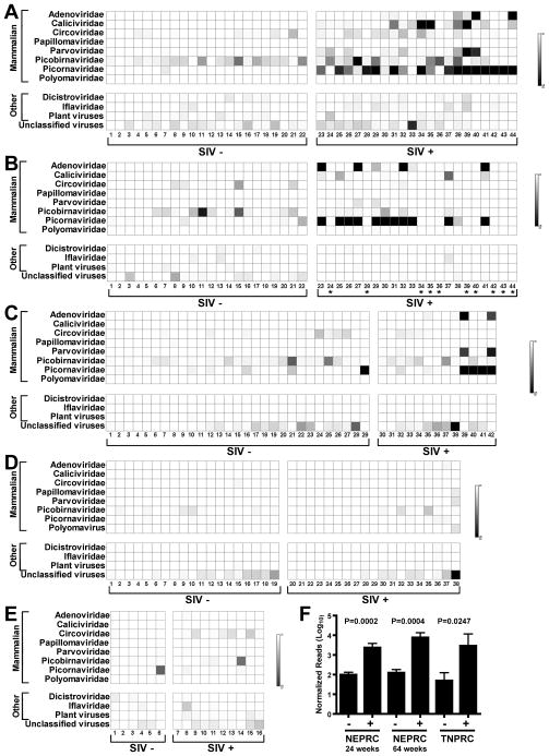
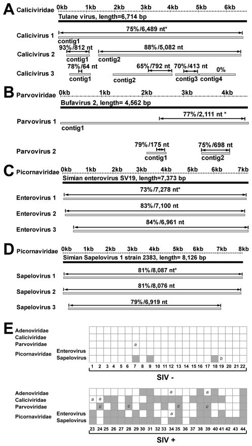
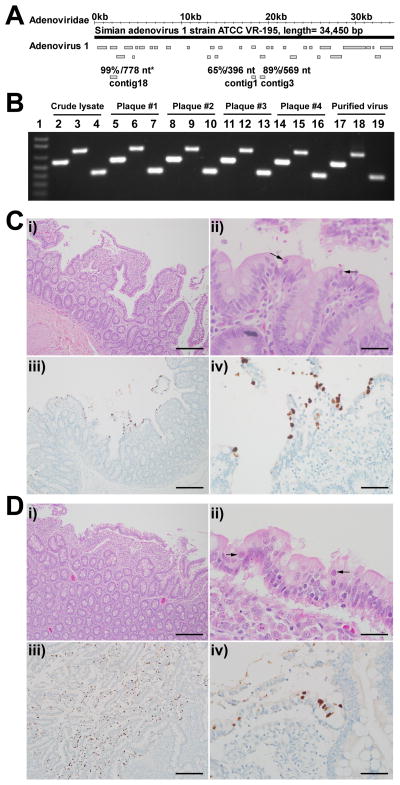
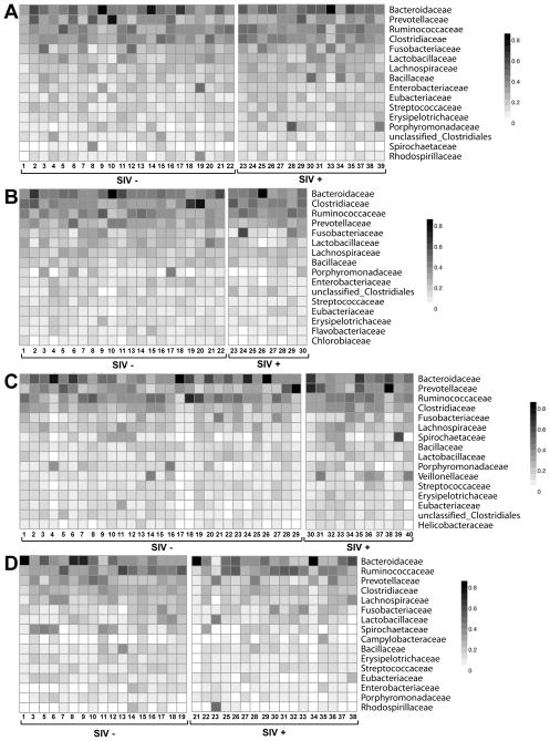
Pathogenic simian immunodeficiency virus (SIV) infection is associated with enteropathy, which likely contributes to AIDS progression. To identify candidate etiologies for AIDS enteropathy, we used next-generation sequencing to define the enteric virome during SIV infection in nonhuman primates. Pathogenic, but not nonpathogenic, SIV infection was associated with significant expansion of the enteric virome. We identified at least 32 previously undescribed enteric viruses during pathogenic SIV infection and confirmed their presence by using viral culture and PCR testing. We detected unsuspected mucosal adenovirus infection associated with enteritis as well as parvovirus viremia in animals with advanced AIDS, indicating the pathogenic potential of SIV-associated expansion of the enteric virome. No association between pathogenic SIV infection and the family-level taxonomy of enteric bacteria was detected. Thus, enteric viral infections may contribute to AIDS enteropathy and disease progression. These findings underline the importance of metagenomic analysis of the virome for understanding AIDS pathogenesis.


Figures

Figure 1
Figure 1. Taxonomic distribution of sequences identified in feces of SIV-infected (SIV+) and uninfected (SIV−) monkeys
A–E graph the percentage of sequences obtained from fecal samples assigned by MEGAN to the indicated taxonomic groups. X-axis numbers refer to individual animals. (A, B) Sequences from monkeys housed at NEPRC for 24 weeks (A) or 64 weeks (B) after intrarectal infection with SIVmac251. *euthanized for progressive AIDS 24 to 64 weeks after SIV infection. (C) Sequences from monkeys housed at the TNPRC 23–64 weeks after intravaginal infection with SIVmac251. (D) Sequences from SIV-infected and control vervet African green monkeys housed at NIH after intravenous infection with SIVagm90, SIVagmVer1 or after natural infection in the wild. (E) Sequences from sabaeus African green monkeys housed at NEPRC and infected intravenously with SIVagmMJ8 or SIVagm9315BR. (A–E) Flanking doughnut charts display the averaged values per kingdom for SIV+ or SIV- monkeys. See also Figure S2
Figure 2
Figure 2. Distribution of virus sequences in rhesus and African green monkeys
For these charts ‘mammalian’ indicates that sequences were most closely related to viruses that infect mammals. Viruses infecting non-mammals are referred to as ‘other’. ‘Unclassified virus’ includes all unclassified viruses, e.g. Chronic bee paralysis virus, Chimpanzee stool associated circular ssDNA virus, Circovirus-like genome RW-C, Circovirus-like genome CB-A, Rodent stool-associated circular genome virus, etc. (A–E) The numbers below each chart and SIV infection status and panels are as in the Figure 1 legend. (F) Comparison of the mean +/− SEM number of picornavirus sequences, after normalization for analysis using MEGAN, detected in the indicated cohorts of SIV-infected (+) and control (−) rhesus monkeys. See also Table S1
Figure 3
Figure 3. Identification of enteric viruses in rhesus monkeys housed at the NEPRC
For panels A–D grey bars represent assembled viral contigs, while black bars represent the genome of the most closely related known virus. *Indicates percent nucleotide identity over the designated length of the best aligned homologous region (indicated by double headed arrow) compared to the most closely virus genome. Animal numbers are as in Figure 1A. (A) Contigs from WUHARV Caliciviruses 1 (animal 39), 2 (from an animal not included in the cohort), and 3 (animal 39) compared to Tulane calicivirus. Calicivirus 1 contig 1 derived from 879 sequences, length= 6,578 bp; Calicivirus 2 contig 1 derived from 16 sequences, length=812 bp; Calicivirus 2 contig 2 assembled from 120 sequences, length= 5,083 bp; Calicivirus 3 contig 1 assembled from 14 sequences, length= 750 bp; Calicivirus 3 contig 2 assembled from 67 sequences, length= 2,111 bp; Calicivirus 3 contig 3 assembled from 41 sequences, length= 832 bp; Calicivirus 3 contig 4 assembled from 38 sequences, length=1,273 bp. (B) Contigs from WUHARV Parvovirus 1 (animal 39) and 2 (animal 35) compared with the sequence of Bufavirus 2. Parvovirus 1 contig 1 assembled from 375 sequences, length= 4,905 bp; Parvovirus 2 contig 1 representing 1 sequence, length= 470 bp; Parvovirus 2 contig 2 assembled from 6 sequences, length= 690 bp. (C) Contigs from WUHARV Enterovirus 1 (animal 41), 2 (animal 39) and 3 (animal 33) compared with the sequence of Simian enterovirus SV19. Enterovirus 1 assembled from 1,084 sequences, length= 7,273 bp; Enterovirus 2 assembled from 758 sequences, length= 7,128 bp; Enterovirus 3 assembled from 406 sequences, length= 6,962 bp. (D) Contigs from WUHARV Sapelovirus 1 (animal 42), 2 (animal 41) and 3 (animal 37) compared with the sequence of Simian Sapelovirus 1 strain 2383. Sapelovirus 1 assembled from 3,081 sequences, length= 8,059 bp; Sapelovirus 2 assembled from 2,711 sequences, length= 8,025 bp; Sapelovirus 3 assembled from 380 sequences, length= 6,872 bp. (E) A chart showing the presence (grey box) of viral sequences in monkeys housed at NEPRC for 24 weeks as detected by PCR using virus-specific primers (Table S2). Numbers below the chart refer to the animals in Figure 1A. “a” = lack of detection of a virus likely due to the presence of a divergent virus; “b” = lack of detection of a virus for unknown reasons. “c” = detection of virus sequences in serum samples taken at the time of euthanasia for AIDs. See also Figure S3 and Table S2
Figure 4
Figure 4. Enteric disease in SIV-infected rhesus monkeys at necropsy
(A) The convention for showing viral contigs is as in Figure 3. Contigs from WUHARV Adenovirus 1 (animal #40) compared to the known virus Simian adenovirus 1 strain ATCC VR-195. These contigs were assembled from 1308 sequences. (B) A gel showing PCR confirmation of WUHARV adenovirus 1 during amplification, plaque purification and cesium chloride gradient purification. The three PCR products for each sample (lanes 2–19) were derived from primers 4302c3f and 4302c3r, 4302c18f and 4302c18r, and 4302c1f and 4302c 1r respectively (Table S2). Lane 1, MW marker. (C, D) Histopathology (top panels) and adenovirus immunohistochemistry (bottom panels) of the small intestine. Adenovirus infection was associated with villous atrophy and fusion (i) and sloughed epithelial cells that contained intranuclear adenoviral inclusions (ii, arrows). Adenovirus antigen could be localized to villous tip epithelium by immunohistochemistry (brown color of DAB chromagen, Mayer’s counterstain; iii and iv). Scale bars in i, iii = 0.5 mm. Scale bars in ii, iv=200 μm.
Figure 5
Figure 5. Representative bacterial families in rhesus and African green monkey feces
For A–D heat maps display the number of sequences assigned to specific bacterial families for individual animals in each cohort. The nature of SIV infection is as defined in the legend of Figure 1. (A, B) Sequences from pathogenic SIV-infected and control rhesus monkeys housed at the NEPRC (A) 24 weeks and (B) 64 weeks after SIV infection. (C) Sequences from pathogenic SIV-infected and control rhesus monkeys housed at the TNPRC. (D) Sequences from nonpathogenic SIV-infected and control vervet African green monkeys housed at the NIH. See also Figure S4

Comment in

References

    1. Altschul SF, Gish W, Miller W, Myers EW, Lipman DJ. Basic local alignment search tool. JMolBiol. 1990;215:403–410. - PubMed
    1. Bailey C, Mansfield K. Emerging and reemerging infectious diseases of nonhuman primates in the laboratory setting. VetPathol. 2010;47:462–481. - PubMed
    1. Barton ES, White DW, Cathelyn JS, Brett-McClellan KA, Engle M, Diamond MS, Miller VL, Virgin HW. Herpesvirus latency confers symbiotic protection from bacterial infection. Nature. 2007;447:326–329. - PubMed
    1. Bench SR, Hanson TE, Williamson KE, Ghosh D, Radosovich M, Wang K, Wommack KE. Metagenomic characterization of Chesapeake Bay virioplankton. Applied and environmental microbiology. 2007;73:7629–7641. - PMC - PubMed
    1. Bloom SM, Bijanki VN, Nava GM, Sun L, Malvin NP, Donermeyer DL, Dunne WM, Jr, Allen PM, Stappenbeck TS. Commensal Bacteroides species induce colitis in host-genotype-specific fashion in a mouse model of inflammatory bowel disease. Cell Host Microbe. 2011;9:390–403. - PMC - PubMed

Publication types

MeSH terms