. 2012 Nov 30;48(4):547-59.
doi: 10.1016/j.molcel.2012.09.002. Epub 2012 Oct 11.

Gravin is a transitory effector of polo-like kinase 1 during cell division

Affiliations

Gravin is a transitory effector of polo-like kinase 1 during cell division

David A Canton et al. Mol Cell. .

Abstract

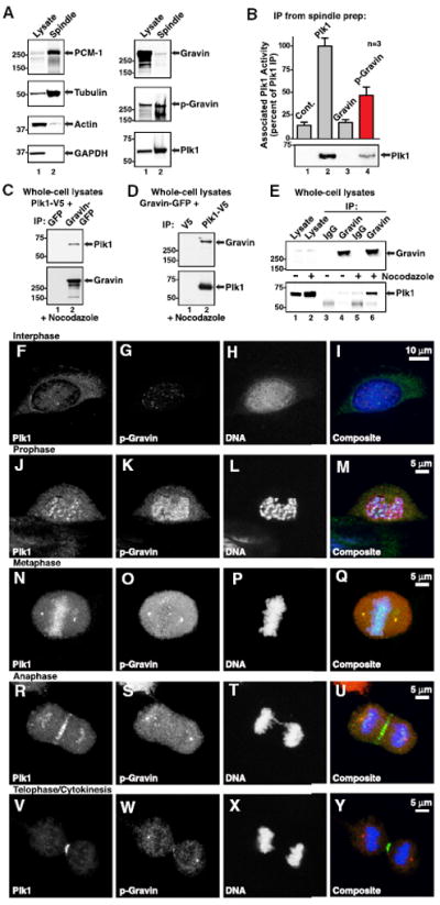
The mitogenic and second-messenger signals that promote cell proliferation often proceed through multienzyme complexes. The kinase-anchoring protein Gravin integrates cAMP and calcium/phospholipid signals at the plasma membrane by sequestering protein kinases A and C with G protein-coupled receptors. In this report we define a role for Gravin as a temporal organizer of phosphorylation-dependent protein-protein interactions during mitosis. Mass spectrometry, molecular, and cellular approaches show that CDK1/Cyclin B1 phosphorylates Gravin on threonine 766 to prime the recruitment of the polo-like kinase Plk1 at defined phases of mitosis. Fluorescent live-cell imaging reveals that cells depleted of Gravin exhibit mitotic defects that include protracted prometaphase and misalignment of chromosomes. Moreover, a Gravin T766A phosphosite mutant that is unable to interact with Plk1 negatively impacts cell proliferation. In situ detection of phospho-T766 Gravin in biopsy sections of human glioblastomas suggests that this phosphorylation event might identify malignant neoplasms.


Figures

Figure 1
Figure 1. Gravin Suppresses Tumor Growth
(A) Immunoblot analysis of U251 cells stably expressing control (lane 1) or Gravin shRNA (lane 2). Staining with antibodies against Gravin (top) and GAPDH (bottom). (B) Densitometric analyses of amalgamated data (n = 3 ± SEM) normalized to GAPDH. (C) Stable U251 cells (3 × 10⁶) were injected into the rear flank of athymic (NU/J) mice. Tumor volume was measured 16–40 days postinjection (black, control shRNA n = 10 ± SEM; red, Gravin shRNA, n = 11 ± SEM; **p ≤ 0.05). (D) Immunoblot analysis of tumor extracts (indicated above each lane) with antibodies against Gravin (top) and GAPDH (bottom). (E) Densitometric analyses of amalgamated data (n = 3 ± SEM). The morphology of paraffin-embedded sections of tumor (F, G, K, and L) was evaluated by H&E staining. Serial sections (H–J, N, and O) costained with anti-Gravin (green) and DRAQ5 (blue) as a nuclear marker confirmed gene silencing of Gravin in situ. (P–R and S–U) Phospho-Histone H3 staining of mitotic cells within the tumor (red). (V) Graph depicting the numbers of phospho-Histone H3-positive cells (per mm2) in control (black) and Gravin shRNA (red) tumor sections (n = 3 individual tumors ± SEM; p = 0.012).
Figure 2
Figure 2. Gravin Is Phosphorylated by CDK1/Cyclin B
Consensus CDK-phosphorylation sites were disrupted by replacing the target serine or threonine with alanine. (A) Gravin mutants (indicated above each lane) were expressed in HEK293 cells. Immunokinase assays were performed on the Flag pull-downs using exogenous CDK1/Cyclin B1 and γ-32P ATP. (B) Quantitation of autoradiographs (n = 3 ± SEM). (C) HEK293 cells were arrested in mitosis with nocodazole (0.1 μg/mL) and treated with the CDK-inhibitor roscovitine (75 μM) in the presence of 32P orthophosphate. Gravin was immunoprecipitated and detected by autoradiography (top) and anti-Gravin immunoblot (bottom). (D) Quantitation of autoradiographs (n = 3 ± SEM). (E) Mass spectrometry analysis of Gravin phosphorylation in vivo; (left panel) all phospho-peptides assigned; (middle panel) phosphosite (human amino acid numbering); (right panel) sites lost upon roscovitine treatment are indicated. (F) Spectral trace of the sequence surrounding phosphothreonine 766. (G) Sequence conservation of threonine 766 (species are indicated). (H) Characterization of phospho-T766 Gravin antiserum on peptide spot arrays (sequences of peptide derivatives are indicated). (I) Gravin was depleted from HEK293 cells with two independent shRNAs. Cell lysates were immunoblotted with antibodies against phospho-T766 Gravin (top), Gravin (middle), and GAPDH (bottom) as a loading control. (J) Gravin was immunoprecipitated from control HEK293 cells or cells arrested in mitosis with nocodazole. Immunoblot detection of phospho-T766 Gravin (top) or Gravin (bottom) is shown. (K) HEK293 cells were arrested in mitosis in the absence (lanes 1 and 2) or presence (lane 3) of roscovitine. Immunoblot detection of phospho-T766 Gravin (top) or Gravin (bottom) in immune complexes is shown.
Figure 3
Figure 3. Phosphorylation of Gravin at Threonine 766 during Mitosis
(A) HEK293 cells were arrested at the G1/S boundary by double thymidine block. Cells were released from the block and harvested every 2 hr from time 0 to 14 (indicated above each lane). Immunoblot detection of phospho-T766 Gravin (top), Gravin (middle) and phospho-S10 Histone H3 (bottom) in cell extracts. (B) Quantitation represents data normalized to GAPDH loading control (phospho-T766 Gravin black trace, phospho-Histone H3 red, Gravin green; n = 3 ± SEM). (C) Immunoblot detection of additional cell-cycle markers as indicated. (D–W) HEK293 cells grown on coverslips were synchronized by double thymidine block, released into fresh media, and fixed in ice-cold methanol 8–9 hr later. Immunofluorescence of cells at defined stages of the cell cycle was performed with antibodies as indicated, followed by detection with secondary antibodies conjugated to Alexa Fluor dyes.
Figure 4
Figure 4. Phospho-T766 Gravin Recruits the Mitotic Kinase Plk1
(A) Spindles were isolated from mitotic cells (Silljé and Nigg, 2006), resuspended in sample buffer, and immunoblotted with specific antibodies as indicated. (B) Mitotic spindles solubilized in lysis buffer were immunoprecipitated with antibodies against Plk1, Gravin, or phospho-T766 Gravin. Immune complexes were subjected to an immunokinase assay using the Plk1 selective peptide -ISDELMDATFADQEAKKK- in the presence of γ-32P-ATP. The relative levels of Plk1 activity are indicated. (C) HEK293 cells were transfected with Plk1-V5 and Gravin-GFP or GFP alone. Copurification of Plk1-V5 (top) and Gravin (bottom) was confirmed by immunoblot. (D) Reciprocal experiments detected copurification of GFP-Gravin (top) in Plk1-V5 (bottom) immune complexes. (E) Immunoblot detection of endogenous Gravin (top) and copurification of Plk1 (bottom) from HEK293 cells. Experiments with control (mIgG) antibodies are included. (F–Y) HEK293 cells on coverslips were synchronized by double thymidine block, released into fresh media, and fixed 8–9 hr later. Immunofluorescence of cells at defined stages of the cell cycle was performed with antibodies against Plk1 (left), phospho-T766 Gravin (middle, left), DNA (middle, right), and composite images (right).
Figure 5
Figure 5. Gravin-Plk1 Interaction Facilitates Cell-Cycle Progression
(A) Mitotic HEK293 cells were treated for 4 hr with H89 (10 μM), Roscovitine (75 μM), Bisindolylmaleimide-1 (100 nM), or GW843682 (10 μM). Gravin immune complexes from each treatment (indicated above each lane) were incubated with a purified recombinant PBD fragment of Plk1 (371–603 aa). PBD-binding was assessed by immunoblot. (B) Quantitation of the data shown in (A) (n = 3 ± SEM). (C) Gravin immune complexes phosphorylated with Plk1 or CDK1/Cyclin B1 were assessed for binding to the recombinant PBD fragment. (D) Quantitation of the data in (C) (n = 3 ± SEM). (E) Flag-tagged Gravin or phosphosite mutants (indicated above each lane) were immunoprecipitated from mitotic cell lysates and incubated with recombinant PBD. Immunoblot detection of Gravin (top) and the PBD fragment (bottom) is shown. (F) Quantitation of the data shown in (E) (n = 3 ± SEM). (G and H) HeLa cells stably expressing Histone 2B-GFP (green) and selected for stable incorporation of control or Gravin shRNAs were transfected with Plk1-Cherry (red). Montage of 633 images at defined times (indicated in each panel) after the cells were released from a double thymidine block. (I) The number of mitotic defects was scored per 100 cells (Control shRNA, n = 12 fields; Gravin shRNA, n = 14 fields; mean ± SEM). (J) Spindle stage profile of HeLa cells expressing control shRNA (light gray; n = 800 ± SEM), Gravin shRNA (red; n = 600 ± SEM), or Gravin shRNA rescued with Gravin-mCherry (dark gray; n = 500 ± SEM). (K) Fluorescence in situ hybridization of Hct116 cells transfected with control and Gravin shRNA. Nuclei were scored as greater than diploid, diploid, and less then diploid using a probe against chromosome 18 (n = 10 fields, >700 nuclei; representative images shown). (L) Soft agar proliferation assay of prostate cancer (PC-3) cells (plated at 10,000 and 5,000 cells in 96-well dishes) expressing control or two independent Gravin shRNAs. Final cells number was analyzed after 6 days using a CyQuant green standard curve (Cell Biolabs; n = 4 ± SEM). (M) Stable PC-3 cell lines were generated expressing Flag-Gravin or Flag-T766A, or transiently transfected with control or Gravin shRNA. Cells were plated in 96-well dishes, and proliferation was analyzed every 3 days by an MTS assay (n = 4 ± SEM, quadruplicate determinations; *p ≤ 0.05).
Figure 6
Figure 6. Phospho-T766 Gravin and Glioblastoma Multiforme
(A) Sagittal section of a brain collected during autopsy exhibiting glioblastoma multiforme (GBM) in the right parietal lobe. (B) Quantitative PCR of Gravin mRNA performed on tissue (white boxes) extracted from the GBM and uninvolved (“healthy”) contralateral side of the brain. Representative Ct trace (left) and Gravin mRNA levels normalized to GAPDH (right), n = 3 ± SEM. (C) Control (IgG) or Gravin immunoprecipitations were performed on brain extracts and probed for reactivity with phospho-T766 Gravin antisera. (D) Magnetic resonance imaging (MRI) of a healthy brain. (E and G) Paraffin-embedded sections of healthy brain tissue stained with H&E. (F) Serial sections costained with antibodies against phospho-S10 Histone H3, phospho-T766 Gravin, and DAPI followed by detection with secondary antibodies conjugated to Alexa Fluor dyes. (H) Additional sections costained with antibodies against Plk1, phospho-T766 Gravin, and DAPI. (I) Magnetic resonance imaging of a glioblastoma localized to the right hemisphere. (J and L) Paraffin-embedded sections of glioblastoma tissue stained with H&E. (K) Equivalent sections costained with antibodies against phospho-S10 Histone H3, phospho-T766 Gravin, and DAPI. (M) GBM sections costained with antibodies against Plk1, phospho-T766 Gravin, and DAPI. (N) The region in (M) indicated by the while box was imaged at higher magnification to visualize costaining of Plk1, phospho-T766 Gravin, and DAPI in mitotic tumor cells. (O) Quantitation of phospho-T766 Gravin-positive cells (per mm2) in sections from control and glioblastoma patients (Control patients, n = 4, 90 fields of view ± SEM; GBM patients n = 4, 90 fields of view ± SEM; p = 0.024).

References

    1. Ahn NG. PORE-ing over ERK substrates. Nat Struct Mol Biol. 2009;16:1004–1005. - PubMed
    1. Akakura S, Huang C, Nelson PJ, Foster B, Gelman IH. Loss of the SSeCKS/Gravin/AKAP12 gene results in prostatic hyperplasia. Cancer Res. 2008;68:5096–5103. - PMC - PubMed
    1. Alexander J, Lim D, Joughin BA, Hegemann B, Hutchins JR, Ehrenberger T, Ivins F, Sessa F, Hudecz O, Nigg EA, et al. Spatial exclusivity combined with positive and negative selection of phosphorylation motifs is the basis for context-dependent mitotic signaling. Sci Signal. 2011;4:ra42. http://dx.doi.org/10.1126/scisignal.2001796. - DOI - PMC - PubMed
    1. Bain J, Plater L, Elliott M, Shpiro N, Hastie CJ, McLauchlan H, Klevernic I, Arthur JS, Alessi DR, Cohen P. The selectivity of protein kinase inhibitors: a further update. Biochem J. 2007;408:297–315. - PMC - PubMed
    1. Brennan IM, Peters U, Kapoor TM, Straight AF. Polo-like kinase controls vertebrate spindle elongation and cytokinesis. PLoS ONE. 2007;2:e409. http://dx.doi.org/10.1371/journal.pone.0000409. - DOI - PMC - PubMed

Publication types

LinkOut - more resources