. 2012 Sep;4(9):648-59.
doi: 10.18632/aging.100490.

Increased IL-21 secretion by aged CD4+T cells is associated with prolonged STAT-4 activation and CMV seropositivity

Affiliations

Increased IL-21 secretion by aged CD4+T cells is associated with prolonged STAT-4 activation and CMV seropositivity

Anshu Agrawal et al. Aging (Albany NY). 2012 Sep.

Abstract

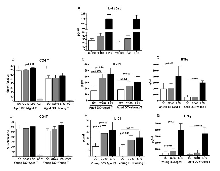
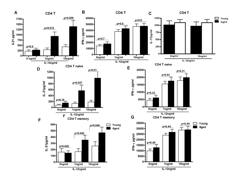
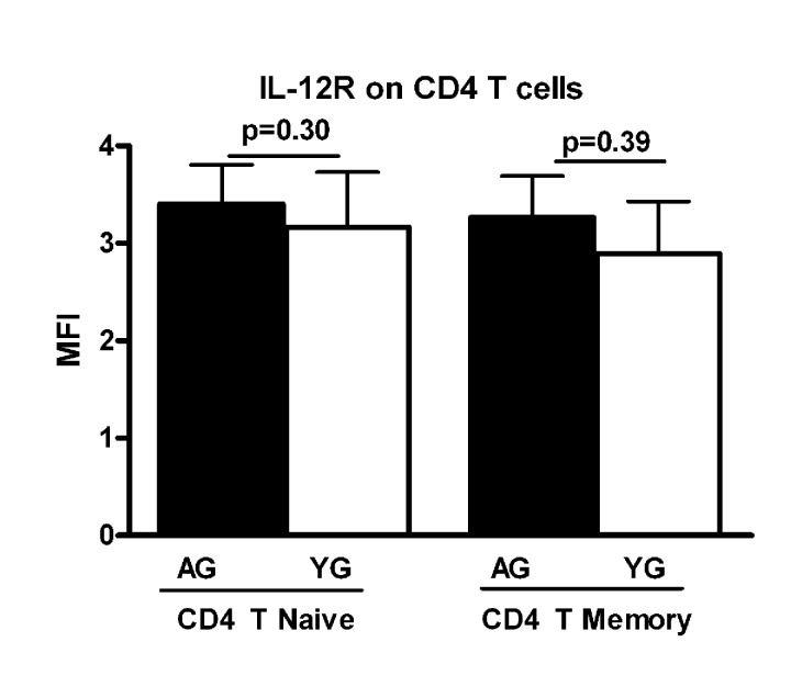
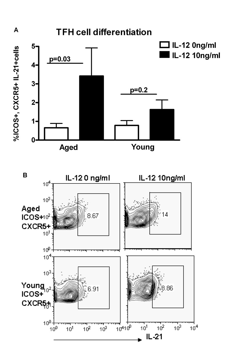
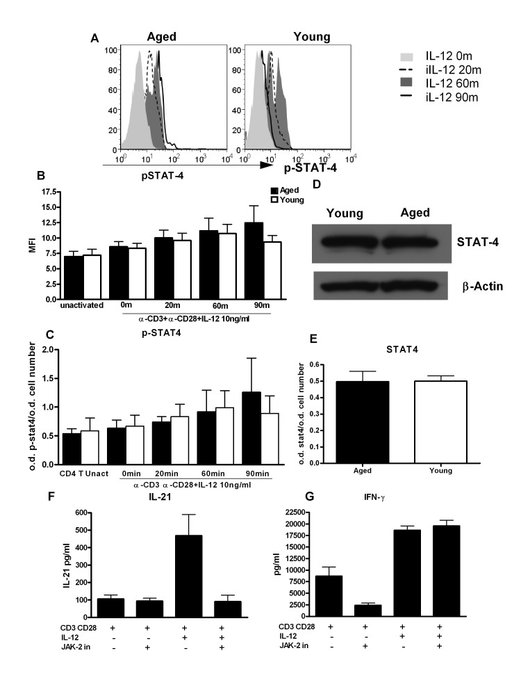
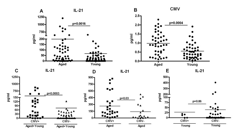
Advancing age leads to significant decline in immune functions. IL-21 is produced primarily by T follicular helper (Tfh) cells and is required for effective immune cell functions. Here we compared the induction of IL-21 in aged and young subjects. Our investigation demonstrates that CD4+T cells from healthy elderly individuals (age ≥ 65) secreted significantly higher levels of IL-21 on priming with aged and young dendritic cells (DC). Though the aged and young DCs secreted comparable levels of IL-12 on stimulation with anti-CD40 antibody and LPS, culture of DCs with aged CD4+ T cells resulted in increased production of IL-21 as compared to that with young CD4+ T cells. Further examination revealed that the response of aged naïve CD4+ T cells to IL-12 was altered, resulting in increased differentiation of aged Th cells towards Tfh cells. Investigation into the signaling mechanism suggested that phosphorylation of STAT-4 in response to IL-12 was sustained for a longer duration in aged CD4+ T cells as compared to CD4+ T cells from young subjects. Additional analysis demonstrated that increased IL-21 secretion correlated with chronic CMV infection in aged subjects. These findings indicate that chronic CMV infection alters the response of aged CD4+ T cells to IL-12 resulting in an increased secretion of IL-21 and that aging affects Tfh cell responses in humans which may contribute to age-associated inflammation and immune dysfunctions.


Conflict of interest statement

The authors of this manuscript have no conflict of interests to declare.

Figures

Figure 1
Figure 1. Increased IL-21 secretion from aged subjects is not due to age-associated alteration in dendritic cell function
A. Bar graph depicts the levels of IL-12p70 in the supernatant from stimulated aged and young DC. B. Bar graph depicts the percent proliferation of aged and young CD4+ T cells after culture with aged DCs. C. Bar graph depicts the level of IL-21 in the supernatant of aged and young CD4+ T cells after culture with aged DCs. D. Bar graph depicts the level of IFN-g in the supernatant of aged and young CD4+ T cells after culture with aged DCs. E. Bar graph depicts the percent proliferation of aged and young CD4+ T cells after culture with young DCs. F. Bar graph depicts the level of IL-21 in the supernatant of aged and young CD4+ T cells after culture with young DCs. G. Bar graph depicts the level of IFN-γ in the supernatant of aged and young CD4+ T cells after culture with young DCs. Data is mean +/− S.E. of 8 different aged and young subjects.
Figure 2
Figure 2. IL-21 secretion is increased from CD4+ T cells from aged subjects
A, B, C Bar graph depicts the levels of IL-21 (A), IFN-γ (B), IL-17 (C) in the supernatant from aged and young total CD4+ T cells after stimulation with IL-12 for 5 days. Data is mean +/− S.E. of 23 different aged and young subjects. D, F. Bar graph depicts the levels of IL-21 secreted from aged and young, Naïve CD4+ T cells (D) and memory CD4+ T cells (F) after stimulation with IL-12 for 5 days. E, G. Bar graph depicts the levels of IL-21 secreted from aged and young, Naïve CD4+ T cells (E) and memory CD4+ T cells (G) after stimulation with IL-12 for 5 days. Data is mean +/− S.E. of 15 different aged and young subjects.
Figure 3
Figure 3. Expression of IL-12R is comparable on aged and young subjects
Bar graph depicts the mean fluorescence intensity (MFI) of IL-12 receptor on naïve and memory CD4+ T cells from aged and young subjects. Data is mean +/− S.E. of 15 different aged and young subjects.
Figure 4
Figure 4. Increased differentiation to T follicular helper cells (Tfh) in aging subjects
A. Bar diagram depicts the percentage of ICOS+, CXCR5+ IL-21+Tfh cells in aged and young CD4+ T cells after activation with IL-12 for 5 days. Data is mean +/− S.E. of 10 different aged and young subjects. B. Contour plot depicts the percent of ICOS+, CXCR5+ IL-21+ Tfh cells in aged and young CD4+ T cells after activation with IL-12 for 5 days. Data is representative of 10 such experiments.
Figure 5
Figure 5. STAT-4 phosphorylation is altered in CD4+ T cells from aged subjects
A. Histograms depict the phosphorylation of STAT-4 in naïve CD4+ T cells from aged and young at 0, 20, 60and 90 m after stimulation with IL-12. Graph is representative of 12 such experiments. B. Bar graph depicts the mean fluorescence intensity (MFI) of phosphorylation of STAT-4 in aged and young CD4+ T cells before and after activation with IL-12. Data is mean +/− S.E. of 12 different aged and young subjects. C. Bar graph also depicts phospho STAT-4 levels, the same as B using in cell ELISA. D. Western blot represents the level of non-phosphorylated STAT-4 in aged and young CD4+ T cells. Data represents pooled samples from 5 different aged and young subjects. E. Bar graph also depicts total STAT as in D using in cell ELISA. F. Graph depicts the level of IL-21 after treatment with JAK-2 inhibitor in aged CD4+ T cells. G. Graph depicts the level of IFN-γ after treatment with JAK-2 inhibitor in aged CD4+ T cells. Data is mean +/− S.E. of 6 different aged subjects.
Figure 6
Figure 6. Increased IL-21 secretion in aged subjects correlates with Cytomegalovirus (CMV) seropositivity
A. Dot blot depicts the level of IL-21 in aged and young plasma. Data is mean of 60 aged and 60 young subjects. Each dot represents one different subject. B. Dot blot depicts the level of CMV seropositivity in aged and young plasma. Data is mean of 50 aged and 50 young subjects. Each dot represents one different subject. C. Dot blot depicts the level of IL-21 in plasma of CMV+ and CMV- individuals. Data is mean of 38 CMV+ and 48 CMV- subjects. Each dot represents one different subject. D. Dot blot depicts the level of IL-21 in plasma of CMV+ and CMV- aged individuals. Data is mean of 32 CMV+ and 18 CMV- aged subjects. Each dot represents one different subject. E. Dot blot depicts the level of IL-21 in plasma of CMV+ and CMV- young individuals. Data is mean of 6 CMV+ and 30 CMV- young subjects. Each dot represents one different subject.

References

    1. Gavazzi G, Herrmann F, Krause KH. Aging and infectious diseases in the developing world. Clin Infect Dis. 2004;39:83–91. - PubMed
    1. McElhaney JE, Effros RB. Immunosenescence: what does it mean to health outcomes in older adults? Curr Opin Immunol. 2009;21:418–424. - PMC - PubMed
    1. Goodwin K, Viboud C, Simonsen L. Antibody response to influenza vaccination in the elderly: a quantitative review. Vaccine. 2006;24:1159–1169. - PubMed
    1. Haynes L. How vaccines work on the background of the aging immune system. Exp Gerontol. 2007;42:438–440. - PubMed
    1. Chen WH, Kozlovsky BF, Effros RB, Grubeck-Loebenstein B, Edelman R, Sztein MB. Vaccination in the elderly: an immunological perspective. Trends Immunol. 2009;30:351–359. - PMC - PubMed

MeSH terms