. 2012 Dec;153(12):5782-95.
doi: 10.1210/en.2012-1595. Epub 2012 Oct 12.

A major lineage of enteroendocrine cells coexpress CCK, secretin, GIP, GLP-1, PYY, and neurotensin but not somatostatin

Affiliations

A major lineage of enteroendocrine cells coexpress CCK, secretin, GIP, GLP-1, PYY, and neurotensin but not somatostatin

Kristoffer L Egerod et al. Endocrinology. 2012 Dec.

Abstract

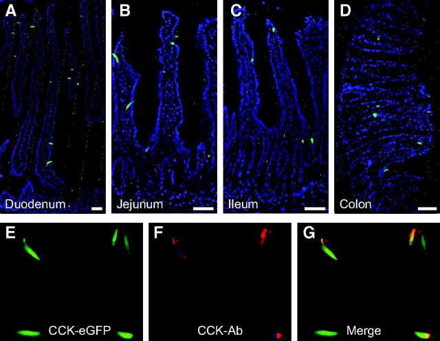
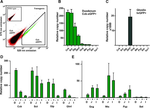
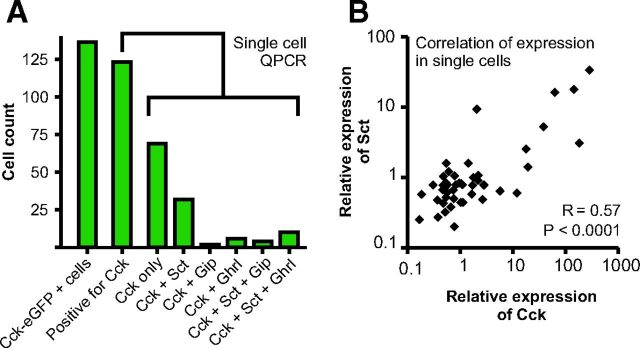
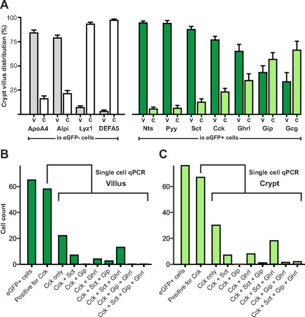
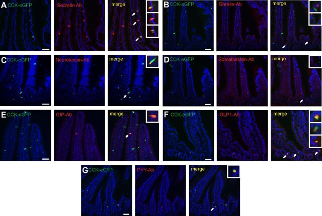
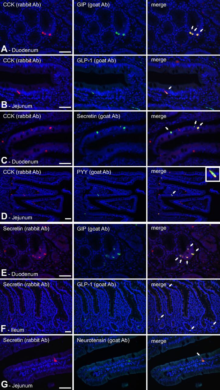
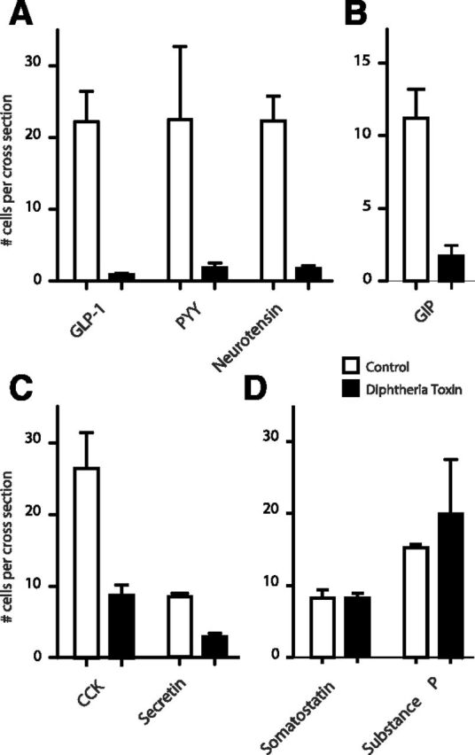
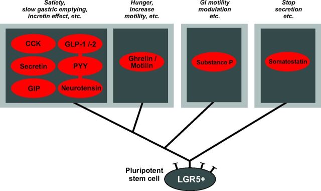
Enteroendocrine cells such as duodenal cholecystokinin (CCK cells) are generally thought to be confined to certain segments of the gastrointestinal (GI) tract and to store and release peptides derived from only a single peptide precursor. In the current study, however, transgenic mice expressing enhanced green fluorescent protein (eGFP) under the control of the CCK promoter demonstrated a distribution pattern of CCK-eGFP positive cells that extended throughout the intestine. Quantitative PCR and liquid chromatography-mass spectrometry proteomic analyses of isolated, FACS-purified CCK-eGFP-positive cells demonstrated expression of not only CCK but also glucagon-like peptide 1 (GLP-1), gastric inhibitory peptide (GIP), peptide YY (PYY), neurotensin, and secretin, but not somatostatin. Immunohistochemistry confirmed this expression pattern. The broad coexpression phenomenon was observed both in crypts and villi as demonstrated by immunohistochemistry and FACS analysis of separated cell populations. Single-cell quantitative PCR indicated that approximately half of the duodenal CCK-eGFP cells express one peptide precursor in addition to CCK, whereas an additional smaller fraction expresses two peptide precursors in addition to CCK. The coexpression pattern was further confirmed through a cell ablation study based on expression of the human diphtheria toxin receptor under the control of the proglucagon promoter, in which activation of the receptor resulted in a marked reduction not only in GLP-1 cells, but also PYY, neurotensin, GIP, CCK, and secretin cells, whereas somatostatin cells were spared. Key elements of the coexpression pattern were confirmed by immunohistochemical double staining in human small intestine. It is concluded that a lineage of mature enteroendocrine cells have the ability to coexpress members of a group of functionally related peptides: CCK, secretin, GIP, GLP-1, PYY, and neurotensin, suggesting a potential therapeutic target for the treatment and prevention of diabetes and obesity.


Figures

Fig. 1.
Fig. 1.
Distribution of eGFP-positive enteroendocrine cells in the GI tract of CCK-eGFP transgenic mice. Panels A–D demonstrate localization of CCK promoter-driven eGFP fluorescence (green) within duodenal (A), jejunal (B), ileal (C), and colonic (D) mucosa along its full glandular extent from the crypts (bottom) to the villus tips. General architecture of the mucosa is visualized by counterstaining with Hoechst nuclei staining (blue). Panels E–G demonstrate localization of CCK promoter-driven eGFP fluorescence (E) with CCK immunoreactivity (F) and merged (G) in jejunum. Bar, 50 μm. Ab, Antibody.
Fig. 2.
Fig. 2.
QPCR analysis of RNAs encoding peptide precursors in isolated, FACS-purified CCK-eGFP positive cells. Panel A, A representative FACS diagram showing the gate (black line surrounding green cells) used for sorting of the CCK-eGFP-positive cells from duodenum based on the green (529 nm) vs. yellow (575 nm) emission after excitation at 488 nm. Panels B and C show relative expression of Cck, Sct, Gip, Ghrl, Gcg, Nts, Pyy, Sst, and Tac. In CCK-eGFP-positive cells from duodenum (B) and ghrelin-hrGFP-positive cells isolated from stomach (C). Panel D shows the expression of the abundant peptide precursor RNAs Cck, Sct, Gip, and Ghrl in CCK-eGFP-positive cells from the duodenum (D) jejunum (J), and ileum (I), respectively. Panel E shows the expression of the scarce peptide precursor RNAs Gcg, Nts, Pyy and Sst in CCK-eGFP-positive cells from the duodenum (D), jejunum (J), and ileum (I), respectively.
Fig. 3.
Fig. 3.
Single-cell QPCR of the four most abundant peptide precursor RNAs (see Fig. 2) Cck, Sct, Gip, and Ghrl in FACS-sorted single CCK-eGFP-positive cells from duodenum. Panel A, In the unamplified QPCR analysis a Ct value less than 40 is considered to be positive for expression of the peptide precursor RNA (see Materials and Methods). The expression pattern for the 123 of the 135 CCK-eGFP-positive cells (far left column) which express Cck itself are shown in the green columns. Of the remaining 12 cells in which Cck expression could not be detected, five expressed Sct, four Ghrl, two Gip, and one both Sct and Ghrl. Panel B, Correlation of the relative expression of Cck and Sct in individual single cells, R = 0.57 (P < 0.0001), using nonparametric Spearman correlation analysis.
Fig. 4.
Fig. 4.
Relative expression of RNAs encoding peptide precursors in FACS-sorted populations of CCK-eGFP-positive cells isolated from villus (V) vs. crypt (C) fractions from duodenum of CCK-eGFP transgenic mice. Populations of cells derived from villus vs. crypts were obtained by collecting cells early vs. late, respectively, during the enzymatic release of cells from the duodenal mucosa (see Material and Methods for details) Panel A (to the left), The relative expression of markers for intestinal villus (Alpi and Apoa4 from enterocytes) and crypt (Lyz1 and Defa5 from Paneth cells) in the CCK-eGFP-negative cells derived from villus (V, gray columns) vs. crypt (C, white columns). Panel A (to the right), The relative expression for the gut hormones: Nts, Pyy, Sct, Cck, Ghrl, Gip, and glucagon/GLP-1 (Gcg) in CCK-eGFP-positive cells derived from villus (V, dark green) vs. crypt (C, light green). The gene expression is indicated as percentage of total, i.e. crypt plus villus. It should be noted that although the actual total expression in the CCK-eGFP-positive cells is relatively high for Cck, Sct, Gip, and Ghrl it is relatively low for Gcg, Pyy, and Nts (see Fig. 2B) the total is here indicated as 100%. Panel B and C, Single-cell QPCR (see legend to Fig. 3) of cells derived from the villus and crypt, respectively. The numbers of cells positive for Cck, Sct, Gip, and ghrelin are given, in this unamplified QPCR analysis a Ct value <40 is considered to be positive for expression of the peptide precursor RNA (see Materials and Methods).
Fig. 5.
Fig. 5.
Immuohistochemical colocalization of selected hormones with CCK-eGFP in duodenum of transgenic CCK-eGFP mice. Vertical panel I, Cck-eGFP-expressing cells (green). Vertical panel II, Immunohistochemical localization of hormones (red) using primary antibodies against secretin (horizontal lane A), ghrelin (lane B), neurotensin (lane C), somatostatin (lane D), gastric inhibitory polypeptide (GIP) (lane E), GLP-1 (lane F), and peptide YY (PYY) (lane G). Vertical panel III, Merged pictures showing colocalization (yellow). Arrows point to colocalization (or, in the case of ghrelin and somatostatin, lack of colocalization) shown in higher magnification in the insets at the top right corner of each panel. Cells storing GLP-1, PYY, and neurotensin show a high degree of colocalization with CCK-eGFP. Secretin- and GIP-producing cells display an approximately 50–60% colocalization with CCK-eGFP. In contrast, somatostatin and ghrelin do not colocalize with CCK-eGFP. Nuclei are visualized with Hoechst counter staining (blue). Bar, 50 μm. Ab, Antibody.
Fig. 7.
Fig. 7.
Immunohistochemical colocalization of selected gut hormones in human small intestine. Panel A, Colocalization of CCK (red, rabbit Ab) and GIP (green, goat Ab) in human duodenum. Panel B, Colocalization of CCK (red, rabbit Ab) and GLP-1 (green, goat Ab) in human jenunum. Panel C, Colocalization of CCK (red, rabbit Ab) and secretin (green, goat Ab) in human duodenum. Panel D, Colocalization of CCK (red, rabbit Ab) and PYY (green, goat Ab) in human jejunum. Panel E, Colocalization of secretin (red, rabbit Ab) and GIP (green, goat Ab) in human duodenum. Panel F, Colocalization of secretin (red, rabbit Ab) and GLP-1 (green, goat Ab) in human ileum. Panel G, Colocalization of secretin (red, rabbit Ab) and neurotensin (green, goat Ab) in human jejunum. For identification of antibodies see Supplemental Table 2. Hoechst nuclei counterstaining (blue) and bar, 50 μm. Ab, Antibody.
Fig. 6.
Fig. 6.
Diphtheria toxin-mediated ablation of enteroendocrine cells in the ileum of transgenic mice expressing the human diphtheria toxin receptor under the control of the proglucagon promoter. Enteroendocrine cells were stained with antibodies selective for GPL-1, PYY, neurotensin, GIP, CCK, secretin, substance P, and somatostatin and counted as described in Materials and Methods in control animals (n = 2, open bars) and 24 h after treatment with low doses of diphtheria toxin (n = 3, closed bars).
Fig. 8.
Fig. 8.
Schematic overview of proposed main enteroendocrine cell lineages of the small intestine. Based on the results of the present study as well as studies by Roth et al. (40), it is suggested that cells expressing the universally inhibitory peptide somatostatin constitute a separate cell lineage distinct from the cell lineage generating cells expressing the rest of the GI-tract peptide hormones. From the latter lineage are apparently generated enterochromafin cells that express substance P (and 5HT) (40), and cells coexpressing ghrelin and motilin (31) plus a major lineage of enteroendocrine cells that have the potential of expressing the functionally related peptides: CCK, secretin, GIP, GLP-1/-2, PYY, and neurotensin. Some of the major functions of the peptide products are indicated above each cell lineage.

References

    1. Schonhoff SE, Giel-Moloney M, Leiter AB 2004. Minireview: Development and differentiation of gut endocrine cells. Endocrinology 145:2639–2644 - PubMed
    1. Rehfeld JF 1998. The new biology of gastrointestinal hormones. Physiol Rev 78:1087–1108 - PubMed
    1. Miller LJ 2003. Gastrointestinal hormones and receptors. In: , Yamada T, ed. Textbook of gastroenterology. Baltimore: Lippincott Williams & Wilkins; 48–76
    1. Field BC, Chaudhri OB, Bloom SR 2010. Bowels control brain: gut hormones and obesity. Nat Rev Endocrinol 6:444–453 - PubMed
    1. Sundler F 2004. GI tract, general anatomy (cells). In: , Martini L, ed. Encyclopedia of endocrine diseases. New York: Elsevier; 208. –215

Publication types

MeSH terms