. 2012 Nov 14;31(22):4334-47.
doi: 10.1038/emboj.2012.280. Epub 2012 Oct 12.

SAS-6 coiled-coil structure and interaction with SAS-5 suggest a regulatory mechanism in C. elegans centriole assembly

Affiliations

SAS-6 coiled-coil structure and interaction with SAS-5 suggest a regulatory mechanism in C. elegans centriole assembly

Renping Qiao et al. EMBO J. .

Abstract

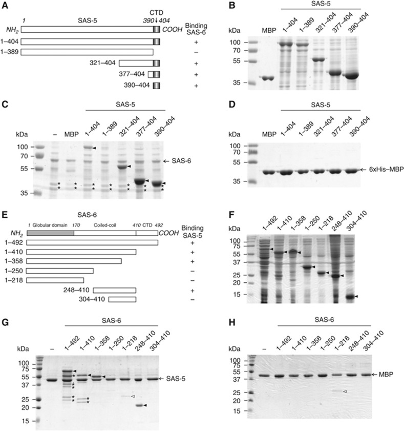
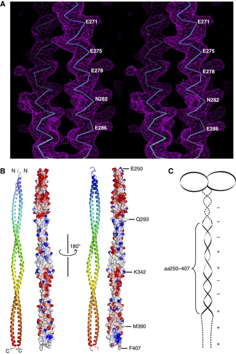
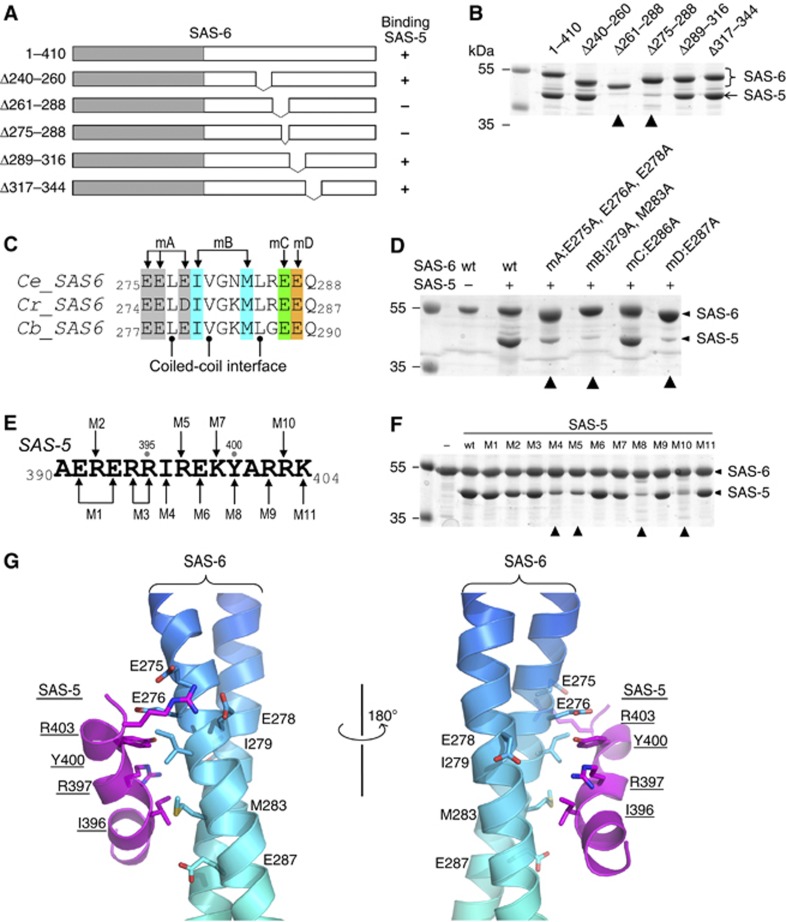
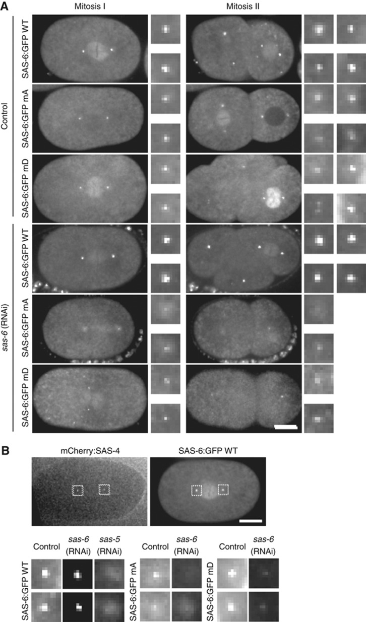
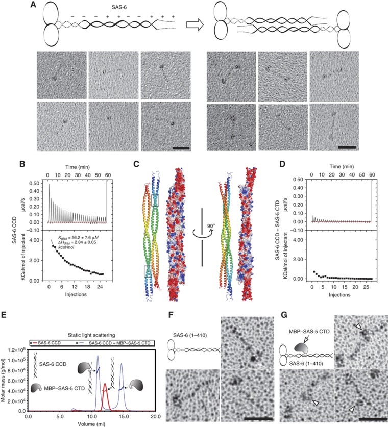
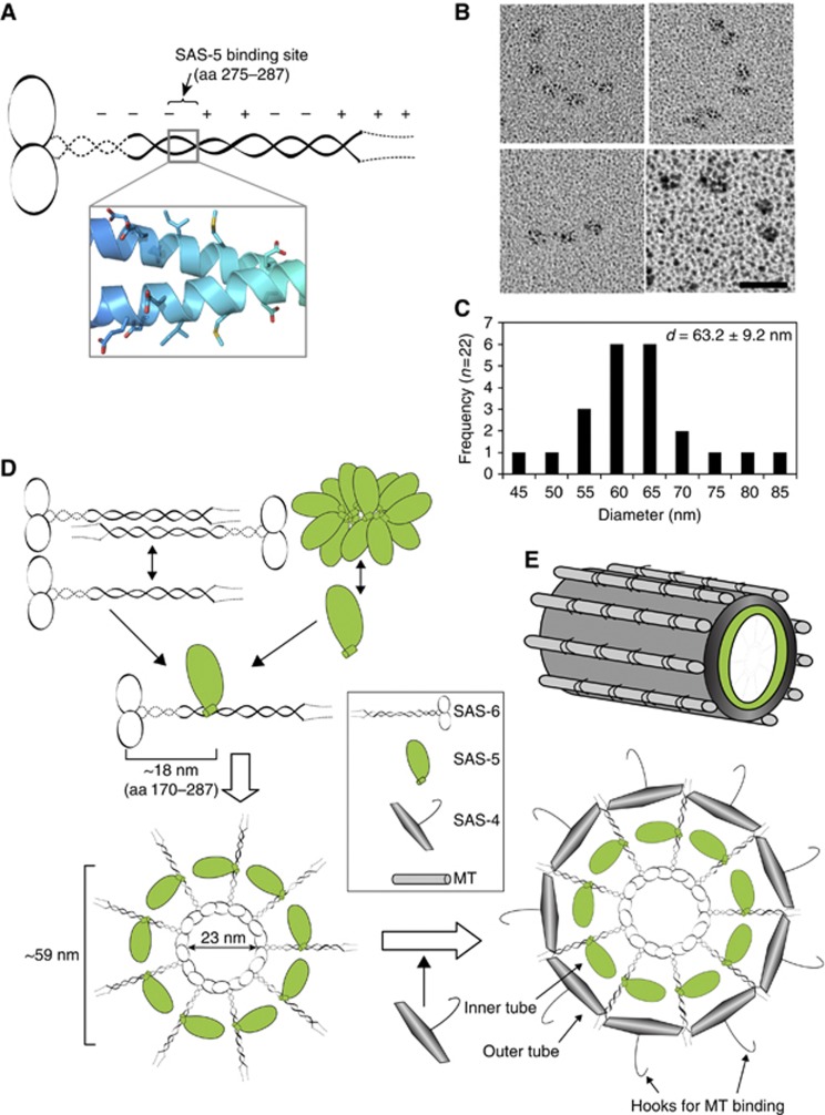
The centriole is a conserved microtubule-based organelle essential for both centrosome formation and cilium biogenesis. Five conserved proteins for centriole duplication have been identified. Two of them, SAS-5 and SAS-6, physically interact with each other and are codependent for their targeting to procentrioles. However, it remains unclear how these two proteins interact at the molecular level. Here, we demonstrate that the short SAS-5 C-terminal domain (residues 390-404) specifically binds to a narrow central region (residues 275-288) of the SAS-6 coiled coil. This was supported by the crystal structure of the SAS-6 coiled-coil domain (CCD), which, together with mutagenesis studies, indicated that the association is mediated by synergistic hydrophobic and electrostatic interactions. The crystal structure also shows a periodic charge pattern along the SAS-6 CCD, which gives rise to an anti-parallel tetramer. Overall, our findings establish the molecular basis of the specific interaction between SAS-5 and SAS-6, and suggest that both proteins individually adopt an oligomeric conformation that is disrupted upon the formation of the hetero-complex to facilitate the correct assembly of the nine-fold symmetric centriole.


Conflict of interest statement

The authors declare that they have no conflict of interest.

Figures

Figure 1
Figure 1
The C-terminal domain of SAS-5 interacts with the central part of the SAS-6 coiled coil. (A) Deletion constructs of SAS-5 used for in vitro binding assays with SAS-6. CTD, carboxy-terminal domain. Numbers indicate amino-acid positions or ranges. The right column shows a summary of the binding results in (C). (B) Purified MBP and soluble fractions of MBP-tagged SAS-5 proteins for in vitro pull-down assays. (C) In vitro pull-down results of SAS-5 proteins using Ni-NTA bound full-length SAS-6 as the bait. MBP is used as a negative control for detecting tag-dependent binding. SAS-5 proteins specifically pulled down by SAS-6 are indicated with arrowheads. Marked by asterisks are the two degradation products of SAS-6. (D) No non-specific interaction to the resin or the MBP tag was detected. (E) Truncation constructs of SAS-6 used for in vitro binding assays with the SAS-5 CTD. The right column shows the summary of the binding results in (G). (F) Soluble fractions of SAS-6 proteins used in the in vitro pull-down assays. Arrowheads indicate the target proteins. (G) In vitro pull-down results of SAS-6 proteins using amylose beads preloaded with the MBP-tagged SAS-5 CTD as the bait. Filled arrowheads indicate SAS-6 proteins pulled down by SAS-5. An empty arrowhead indicates the MBP-dependent non-specific binding of the construct containing residues 1–218 of SAS-6, which is comparable to what is seen in the control experiment in (H). Asterisks indicate the degradation products of SAS-6. (H) Control experiments to show no non-specific binding of SAS-6 proteins to the MBP tag.
Figure 2
Figure 2
Crystal structure of the SAS-6 CCD. (A) Stereo view of a representative portion of the 2FoFc experimental electron density map covering residues 268–290 (contoured at 2.0 σ). For clarity, only the main chains of the final model are shown. (B) Ribbon diagram and electrostatic surface plot of the SAS-6 CCD structure. Residues at the boundaries of differently charged segments are indicated. (C) Schematic representation of the SAS-6 dimer. Dashed lines indicate the regions lacking a known structure. Positive and negative charges along the coiled coil and in the C-terminal domain are depicted as ‘+’ and ‘−’, respectively.
Figure 3
Figure 3
Association of the SAS-5 CTD and the SAS-6 CCD is mediated by synergistic hydrophobic and electrostatic interactions. (A) Schematic of SAS-6 deletion constructs. The right column summarizes the interaction results in (B). (B) In vitro pull-down results of MBP-tagged SAS-5 CTD using Ni-NTA bound SAS-6 as the bait. The two deletions of SAS-6 that failed to pull down SAS-5 are indicated with arrowheads. (C) Sequence alignment of the SAS-5-binding site from three Caenorhabditis species. Ce, Caenorhabditis elegans; Cr, C. remanei; Cb, C. briggsae. Mutations of the four groups of conserved, solvent-exposed residues (to alanines) are highlighted in different colours. (D) Coomassie stained SDS–PAGE gel showing the result of in vitro pull-down of SAS-5 by wild-type (wt) and the four mutations of SAS-6. All mutations except for mC failed to interact with SAS-5. (E) Sequence of the SAS-5 CTD. Eleven mutations are indicated as M1–M11. (F) Coomassie stained SDS–PAGE gel showing the results of in vitro pull-down of wild-type or mutants of the SAS-5 CTD by SAS-6. The four mutations that show a drastic decrease of binding to SAS-5 are indicated with arrowheads. (G) Docking the SAS-5 CTD to its binding site on the SAS-6 CCD by ClusPro 2.0 (Kozakov et al, 2010). Side chains of the residues that participate in the interaction are shown.
Figure 4
Figure 4
In vivo analysis of SAS-6 mutants mA and mD in C. elegans embryos. (A) GFP fusions to wild-type SAS-6 and mutants mA and mD localize to centrioles. However, only wild-type SAS-6:GFP can sustain centriole assembly and consequently spindle bipolarity following depletion of endogenous SAS-6 by isoform-specific RNAi. All wild-type SAS-6:GFP embryos displayed bipolar second divisions following depletion of endogenous SAS-6, all mutant embryos monopolar second divisions. N=10/9 embryos wild-type, 8/22 embryos mA, and 11/17 embryos mD (control/RNAi). Note that GFP signal in RNAi-depleted mutant embryos reflects sperm centriole-associated SAS-6:GFP unaffected by depletion. (B) Centriolar recruitment of SAS-6 requires interaction with SAS-5. Mating was used to introduce mCherry:SAS-4-labelled centrioles into each SAS-6:GFP strain, thereby marking the site of centriole assembly. All embryos are in late prophase-metaphase of the first embryonic division. SAS-6 was detected in wild-type SAS-6:GFP strain following depletion of endogenous SAS-6, but severely diminished in SAS-6 mutants as well as following depletion of SAS-5 in wild-type worms. Scale bar is 10 μm. Insets are magnified × 3. Images in (B) are scaled identically across all strains and conditions to allow cross-comparison.
Figure 5
Figure 5
C. elegans SAS-6 alone forms an anti-parallel tetramer, whereas binding of SAS-5 disrupts the tetrameric association of SAS-6. (A) Schematic model and the rotary metal shadowing electron micrographs of recombinant SAS-6. Scale bars: 30 nm. (B) Experimental and integrated dilution ITC curves for the SAS-6 CCD (residues 248–410). (C) Docking of the SAS-6 CCD self-association by the automated protein docking program ClusPro 2.0 (Kozakov et al, 2010). Both ribbon diagrams and electrostatic surface plots are shown. Boxed are the SAS-5-binding sites on the SAS-6 CCD. (D) Experimental and integrated dilution ITC curves for SAS-6 CCD+SAS-5 CTD. (E) SLS analysis of the complex of the SAS-6 CCD and the MBP–SAS-5 CTD. The SAS-6 CCD by itself forms a dimer (Mw ∼38 kDa), whereas mixing it with the MBP–SAS-5 CTD (molar ratio=1:1.5) gave rise to a hetero-trimer (Mw ∼80 kDa). Mw of the MBP–SAS-5 CTD is 44 kDa. (F) Rotary metal shadowing electron micrographs of SAS-6 (residues 1–410). Scale bar: 30 nm. (G) Rotary shadowing electron micrographs of SAS-6 in complex with MBP-tagged SAS-5 CTD. Arrowheads indicate the bound SAS-5 on the SAS-6 coiled coil. Scale bar: 30 nm.
Figure 6
Figure 6
The SAS-5/SAS-6 complex forms curved structures similar to the central tube of C. elegans centrioles. (A) Schematic of the SAS-6 dimer and the nearly symmetric arrangement of the residues involved in SAS-5 binding. (B) Rotary metal shadowing electron micrographs of the full-length SAS-5-/SAS-6 complex. Scale bar: 30 nm. (C) Histogram representation of mean diameters of the rings measured from circle structures. The majority of the circle structures have a diameter of 60–65 nm, which is in good agreement with that of the central tube of C. elegans centrioles. (D) Hypothetical mechanism of recruitment of SAS-5, SAS-6, and SAS-4. (E) Schematic illustration of the C. elegans centriole. The inner layer of the central tube is formed by SAS-5, whereas the outer layer is built by SAS-4, which has a hook-like structure that serves to recruit the singlet MTs.

References

    1. Andersen JS, Wilkinson CJ, Mayor T, Mortensen P, Nigg EA, Mann M (2003) Proteomic characterization of the human centrosome by protein correlation profiling. Nature 426: 570–574 - PubMed
    1. Arquint C, Sonnen KF, Stierhof YD, Nigg EA (2012) Cell-cycle-regulated expression of STIL controls centriole number in human cells. J Cell Sci 125: 1342–1352 - PubMed
    1. Azimzadeh J, Marshall WF (2010) Building the centriole. Curr Biol 20: R816–R825 - PMC - PubMed
    1. Basto R, Lau J, Vinogradova T, Gardiol A, Woods CG, Khodjakov A, Raff JW (2006) Flies without centrioles. Cell 125: 1375–1386 - PubMed
    1. Battye TG, Kontogiannis L, Johnson O, Powell HR, Leslie AG (2011) iMOSFLM: a new graphical interface for diffraction-image processing with MOSFLM. Acta Crystallogr D Biol Crystallogr 67: 271–281 - PMC - PubMed

Publication types

MeSH terms

Substances

LinkOut - more resources