. 2013 Feb:50:69-75.
doi: 10.1016/j.nbd.2012.10.005. Epub 2012 Oct 12.

Neurodevelopmental impairment following neonatal hyperoxia in the mouse

Affiliations

Neurodevelopmental impairment following neonatal hyperoxia in the mouse

Manimaran Ramani et al. Neurobiol Dis. 2013 Feb.

Abstract

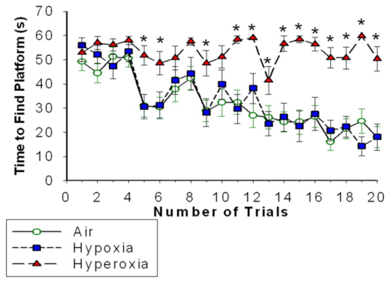
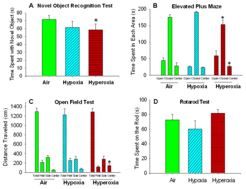
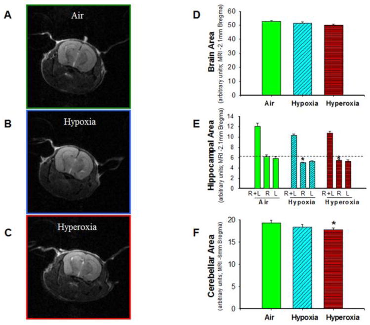
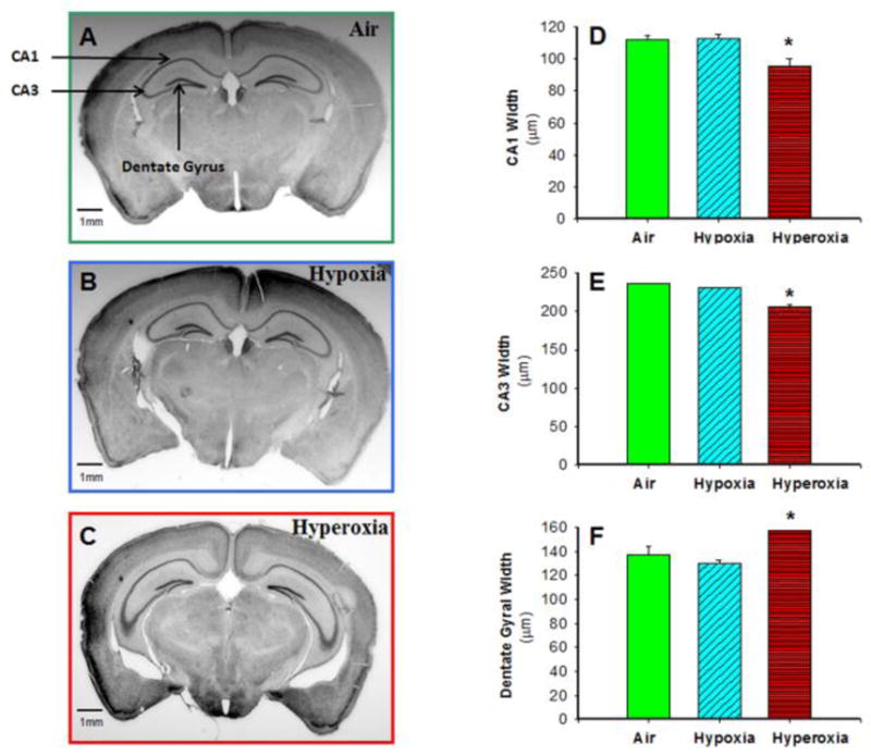
Extremely premature infants are often exposed to supra-physiologic concentrations of oxygen, and frequently have hypoxemic episodes. These preterm infants are at high risk (~40%) for neurodevelopmental impairment (NDI) even in the absence of obvious intracranial pathology such as intraventricular hemorrhage or periventricular leukomalacia. The etiology for NDI has not been determined, and there are no animal models to simulate neurodevelopmental outcomes of prematurity. Our objectives were to develop and characterize a mouse model to determine long-term effects of chronic hypoxia or hyperoxia exposure on neurodevelopment. Newborn C57BL/6 mice were exposed to hypoxia (12% O(2)) or hyperoxia (85% O(2)) from postnatal days 1 to 14 and then returned to air. At 12-14 weeks of age, neurobehavioral assessment (Water Maze test, Novel Object Recognition test, Open Field test, Elevated Plus Maze, and Rotarod test) was performed, followed by MRI and brain histology. Neurobehavioral testing revealed that hyperoxia-exposed mice did poorly on the water maze and novel object recognition tests compared to air-exposed mice. MRI demonstrated smaller hippocampi in hyperoxia- and hypoxia-exposed mice with a greater reduction in hyperoxia-exposed mice, including a smaller cerebellum in hyperoxia-exposed mice. Brain histology showed reduced CA1 and CA3 and increased dentate gyral width in hippocampus. In conclusion, neonatal hyperoxia in mice leads to abnormal neurobehavior, primarily deficits in spatial and recognition memory, associated with smaller hippocampal sizes, similar to findings in ex-preterm infants. This animal model may be useful to determine mechanisms underlying developmental programming of NDI in preterm infants, and for evaluation of therapeutic strategies.


Conflict of interest statement

Conflict of interest: None

Figures

Figure 1
Figure 1. Neonatal hyperoxia exposure decreased adult spatial learning and memory
Time (seconds) taken by the adult mice exposed to Air (line with open green circles), Hypoxia (dashed line with blue squares) and Hyperoxia (dashed line with red triangles) to find the platform in water maze navigation task. Mice exposed to air or hypoxia in the neonatal period performed comparably, but neonatal exposure to hyperoxia impaired the ability of mice to find the submerged platform. means ± SEM; n= 22 in air, 13 in hypoxia, and 16 in hyperoxia.*p<0.05 vs. air-exposed mice RM ANOVA).
Figure 2
Figure 2. Neonatal hyperoxia exposure decreased memory, increased exploratory behavior and decreased anxiety in adult mice
(A) Novel Object Recognition test: Hyperoxia-exposed mice spent less time with novel objects as compared to air-exposed mice. (B) Elevated Plus Maze: Hyperoxia-exposed mice spent more time in the open center and less time in the closed area. (C) Open Field Test: Hyperoxia-exposed mice traveled a greater distance in the center area (D) Rotarod test: No statistical differences were noted in time spent on the rod. Air-exposed: solid green bars, hypoxia-exposed mice: cyan angled stripes, hyperoxia-exposed: red bars with horizontal stripes; means± SEM; n= 10 in air, 7 in hypoxia, and 7 in hyperoxia.*p<0.05 vs. air-exposed mice.
Figure 3
Figure 3. Neonatal hypoxia and hyperoxia exposure decreased hippocampal and cerebellar size in adult mice
T2-weighted non-contrast MR images of brain from air-exposed (A), hypoxia-exposed (B), and hyperoxia-exposed (C) mice were used to measure brain, hippocampal, and cerebellar areas by tracing the periphery of the specific regions. (D) Cross-sectional brain area in adult mice at approximately −2.1 mm bregma did not differ by neonatal exposure to air, hypoxia or hyperoxia. (E) Cross-sectional area of the right hippocampus at −2.1 mm bregma was lower in hypoxia- and hyperoxia-exposed mice, as compared to air-exposed mice. (F) Cross-sectional cerebellar area at −6 mm bregma was reduced in neonatal hyperoxia-exposed mice as compared to air-exposed mice. Air-exposed: solid green bars, hypoxia-exposed mice: cyan angled stripes, hyperoxia-exposed: red bars with horizontal stripes; means± SEM; n= 11 in air, 7 in hypoxia, and 6 in hyperoxia.*p<0.05 vs. air-exposed mice.
Figure 4
Figure 4. Neonatal hyperoxia exposure decreased CA1 and CA3 width but increased dentate gyral width of hippocampus in adult mice
Photomicrographs of Nissl-stained adult mouse brain sections from air-exposed (A), hypoxia-exposed (B), and hyperoxia-exposed (C) mice were used to measure CA1, CA3, and dentate gyral width by image analysis. CA1 width (D) and CA3 width (E) in adult mice at approximately −2.1 mm bregma were lower, but dentate gyral width (F) was increased in adult mice exposed to neonatal hyperoxia, as compared to mice exposed to air or hypoxia. Air-exposed: solid green bars, hypoxia-exposed mice: cyan angled stripes, hyperoxia-exposed: red bars with horizontal stripes; means± SEM; n= 11 in air, 7 in hypoxia, and 6 in hyperoxia.*p<0.05 vs. air-exposed mice. Calibration bar = 1 mm.

References

    1. Aarnoudse-Moens CS, et al. Meta-analysis of neurobehavioral outcomes in very preterm and/or very low birth weight children. Pediatrics. 2009;124:717–28. - PubMed
    1. Ambalavanan N, et al. Endothelin-A receptor blockade prevents and partially reverses neonatal hypoxic pulmonary vascular remodeling. Pediatr Res. 2005;57:631–6. - PMC - PubMed
    1. Ambalavanan N, et al. Transforming growth factor-beta signaling mediates hypoxia-induced pulmonary arterial remodeling and inhibition of alveolar development in newborn mouse lung. Am J Physiol Lung Cell Mol Physiol. 2008a;295:L86–95. - PMC - PubMed
    1. Ambalavanan N, et al. Role of matrix metalloproteinase-2 in newborn mouse lungs under hypoxic conditions. Pediatr Res. 2008b;63:26–32. - PMC - PubMed
    1. Anderson PJ, Doyle LW. Neurodevelopmental outcome of bronchopulmonary dysplasia. Semin Perinatol. 2006;30:227–32. - PubMed

Publication types