Review
. 2013 Jul;70(14):2519-32.
doi: 10.1007/s00018-012-1183-2. Epub 2012 Oct 12.

Intrinsic control of mammalian retinogenesis

Affiliations
Review

Intrinsic control of mammalian retinogenesis

Mengqing Xiang. Cell Mol Life Sci. 2013 Jul.

Abstract

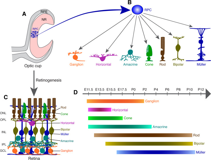
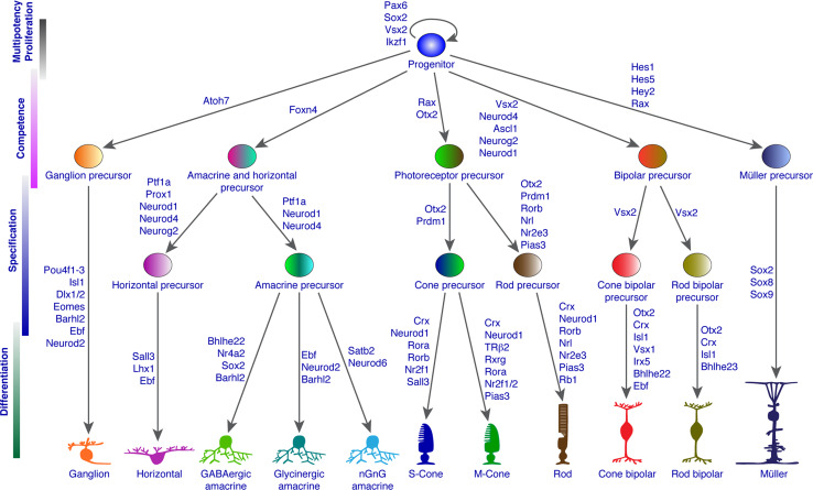
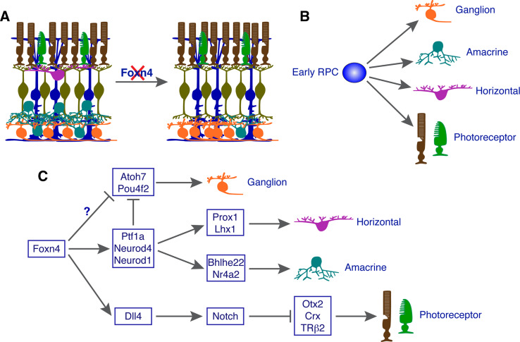
The generation of appropriate and diverse neuronal and glial types and subtypes during development constitutes the critical first step toward assembling functional neural circuits. During mammalian retinogenesis, all seven neuronal and glial cell types present in the adult retina are specified from multipotent progenitors by the combined action of various intrinsic and extrinsic factors. Tremendous progress has been made over the past two decades in uncovering the complex molecular mechanisms that control retinal cell diversification. Molecular genetic studies coupled with bioinformatic approaches have identified numerous transcription factors and cofactors as major intrinsic regulators leading to the establishment of progenitor multipotency and eventual differentiation of various retinal cell types and subtypes. More recently, non-coding RNAs have emerged as another class of intrinsic factors involved in generating retinal cell diversity. These intrinsic regulatory factors are found to act in different developmental processes to establish progenitor multipotency, define progenitor competence, determine cell fates, and/or specify cell types and subtypes.


Figures

Fig. 1
Fig. 1
Retinal development from multipotent progenitor cells. a, b Schematic illustration of the double-layered optic cup. The inner layer harbors multipotent retinal progenitors that are capable of differentiating into the ganglion, horizontal, amacrine, cone, rod, bipolar, and Müller cells. c Schematic of the retinal structure assembled from the seven cell types produced from multipotent progenitors. d Order of birth of mouse retinal cell types. Birthdating analysis has revealed a loose temporal sequence of generation of the six neuronal and one glial cell types in the mouse retina. GCL ganglion cell layer, INL inner nuclear layer, IPL inner plexiform layer, ONL outer nuclear layer, OPL outer plexiform layer, RPC retinal progenitor cell, RPE retinal pigment epithelium, NR neural retina
Fig. 2
Fig. 2
Known transcription factors and cofactors involved in retinal progenitor multipotency and competence as well as in the specification and differentiation of different retinal cell types and subtypes
Fig. 3
Fig. 3
Model by which Foxn4 promotes the amacrine and horizontal cell fates but suppresses the alternative photoreceptor and ganglion cell fates in early retinal progenitor cells (RPCs). a Schematic illustration of retinal phenotype in Foxn4 null mutant mice. b Early RPCs are capable of generating ganglion, amacrine, horizontal, and photoreceptor cells. c Foxn4 specifies early RPCs into amacrine and horizontal cells by activating the expression of Ptf1a, Neurod1, and Neurod4, three bHLH transcription factors (TFs) involved in the specification of these two cell types. Meanwhile, Foxn4 may simultaneously suppress the ganglion fate also by activating the expression of these three bHLH factors due to their activity to repress the expression of Atoh7 and Pou4f2. Another possibility is that Foxn4 may directly repress Atoh7 and Pou4f2 expression. Foxn4 suppresses photoreceptor fates by directly activating Dll4-Notch signaling which in turn represses the expression of Otx2, Crx, TRβ2, and perhaps other TFs involved in photoreceptor fate determination and differentiation

References

    1. Vaney DI. Retinal neurons: cell types and coupled networks. Prog Brain Res. 2002;136:239–254. - PubMed
    1. Wassle H, Boycott BB. Functional architecture of the mammalian retina. Physiol Rev. 1991;71:447–480. - PubMed
    1. Masland RH. The fundamental plan of the retina. Nat Neurosci. 2001;4:877–886. - PubMed
    1. Masland RH. Neuronal diversity in the retina. Curr Opin Neurobiol. 2001;11:431–436. - PubMed
    1. Sidman RL (1961), Histogenesis of mouse retina studied with thymidine-3H. In: Smelser G (ed) The structure of the eye. Academic, New York, pp 487–506

Publication types

MeSH terms

LinkOut - more resources