. 2013 Jan;23(1):46-59.
doi: 10.1101/gr.138842.112. Epub 2012 Oct 11.

Evolutionary dynamism of the primate LRRC37 gene family

Affiliations

Evolutionary dynamism of the primate LRRC37 gene family

Giuliana Giannuzzi et al. Genome Res. 2013 Jan.

Abstract

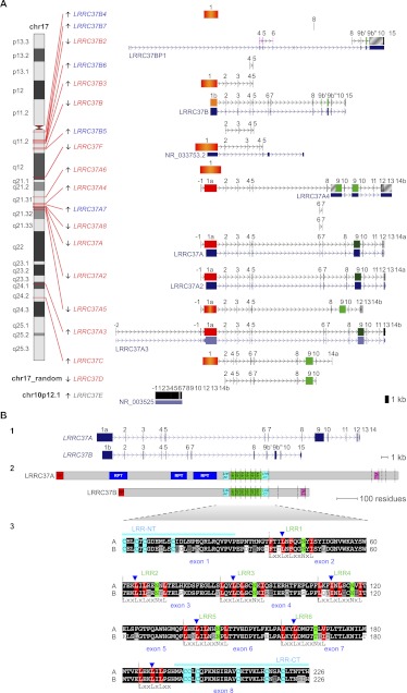
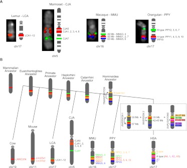
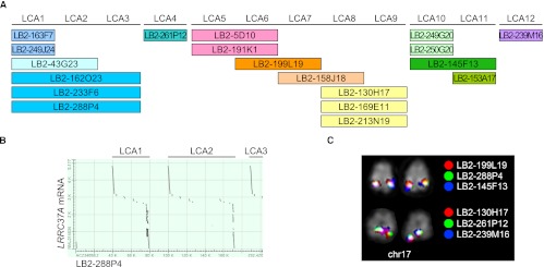
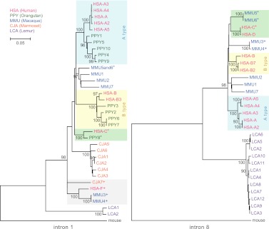
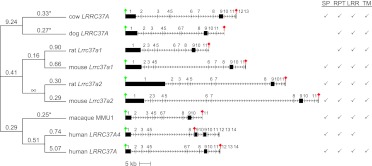
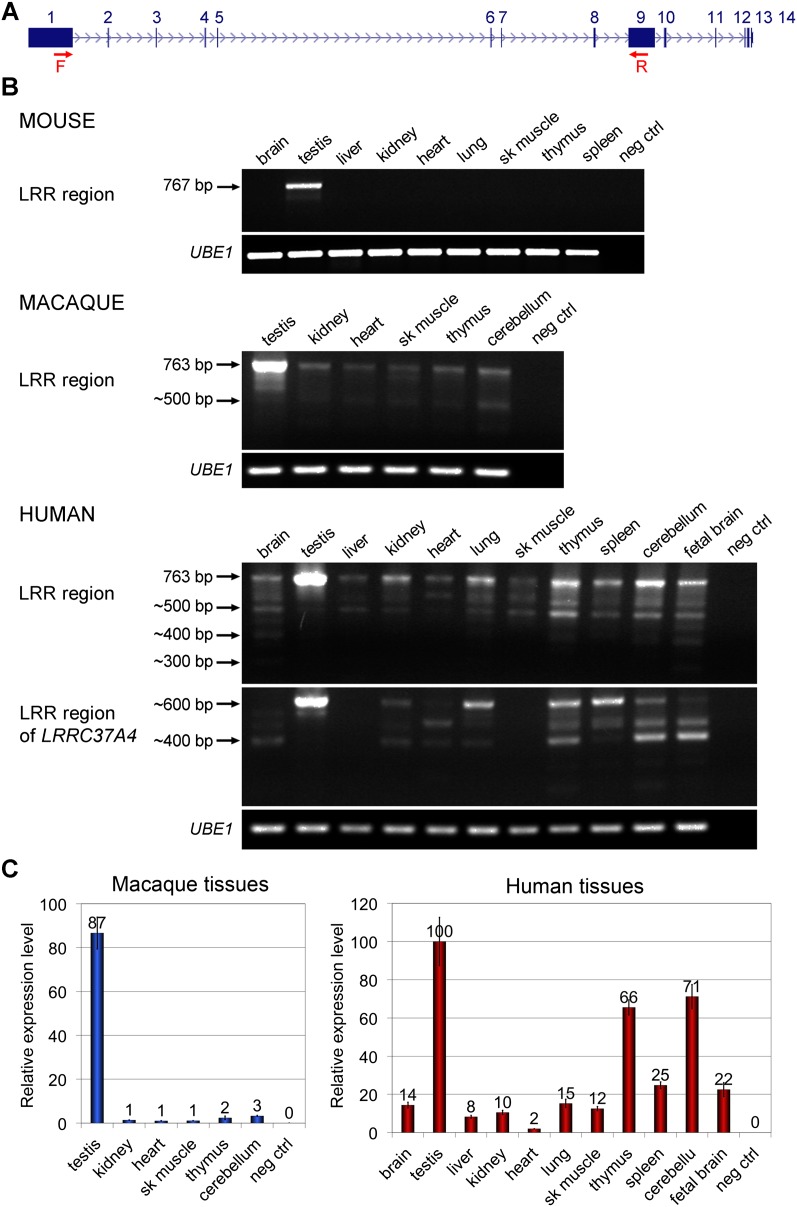
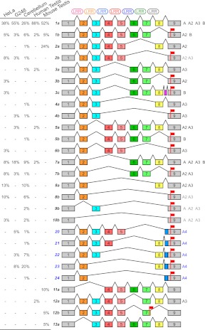
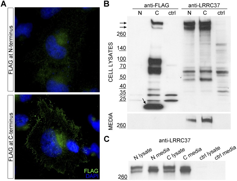
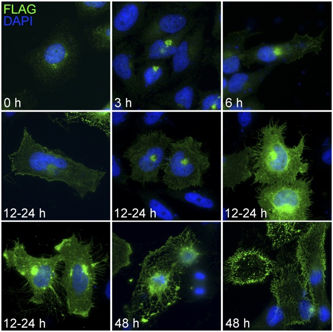
Core duplicons in the human genome represent ancestral duplication modules shared by the majority of intrachromosomal duplication blocks within a given chromosome. These cores are associated with the emergence of novel gene families in the hominoid lineage, but their genomic organization and gene characterization among other primates are largely unknown. Here, we investigate the genomic organization and expression of the core duplicon on chromosome 17 that led to the expansion of LRRC37 during primate evolution. A comparison of the LRRC37 gene family organization in human, orangutan, macaque, marmoset, and lemur genomes shows the presence of both orthologous and species-specific gene copies in all primate lineages. Expression profiling in mouse, macaque, and human tissues reveals that the ancestral expression of LRRC37 was restricted to the testis. In the hominid lineage, the pattern of LRRC37 became increasingly ubiquitous, with significantly higher levels of expression in the cerebellum and thymus, and showed a remarkable diversity of alternative splice forms. Transfection studies in HeLa cells indicate that the human FLAG-tagged recombinant LRRC37 protein is secreted after cleavage of a transmembrane precursor and its overexpression can induce filipodia formation.


Figures

Figure 1.
Figure 1.
(A) LRRC37 family organization in human. The structure of complete and partial LRRC37 genes according to the reference human genome (hg18) is shown. (Left) Chromosomal band location of each locus on human chromosome 17 ideogram (only LRRC37E maps to chromosome 10 [gray] and is a retrocopy; LRRC37D does not have a chromosomal assignment). Genes are indicated as expressed (red) or not expressed (blue) based on the analysis of EST (expressed sequence tag) data. (Right) Variation in LRRC37 structures. Exons are represented as blocks connected by horizontal lines with arrowheads depicting introns. Exon 1 of A, A2, A3, and A4 copies (1a, in red) is longer than exon 1 of the B copy (1b, in orange); the B and B2 copies have exon 9 sequence split into two exons, 9b′ and 9b″. Exon 1 with deletions and/or insertions is red-orange. Exon 9 is dark green; exon 9 with deletions and/or insertions is light green. LRRC37A3 copy has one additional exon located at 5′ but lacks exon 12, and exon 13 is not transcribed. LRRC37A4 copy lacks exons 6 and 7 but carries a tandem duplication of exons 9 and 10. LRRC37A5 has a deletion of a single nucleotide at the end of the exon 1 sequence, causing a frameshift of the reading frame and the formation of a stop codon at the end of exon 1. LRRC37B2 is a fusion gene: exons 7, 8, and 9 match exons 9b′, 9b″, 10, and 15 of LRRC37B, whereas exons 4, 5, and 6 derive from a duplication of exons 4, 5, and 6 of SMAD-specific E3 ubiquitin protein ligase 2 (SMURF2) (NM_022739) (in pink), which maps at 17q24.1. Exapted introns are gray striped. RefSeq genes annotated in these loci are reported, following the same display conventions as in the UCSC Genome Browser. (B) Human LRRC37A and LRRC37B gene and protein structures. (Top panel) LRRC37A and LRRC37B cDNA predict an ORF with the methionine start codon (exon 1) and a stop codon (exon 14 and exon 15, respectively). LRRC37A encodes a predicted protein of 1700 amino acids with a molecular weight of 188 kDa. LRRC37B encodes a predicted protein of 947 amino acids with a molecular weight of 106 kDa. (Middle panel) Predicted structure of human LRRC37A and LRRC37B proteins according to SMART and LRRscan tools. Signal peptides (red), LRR motifs (green), LRR-NT and LRR-CT domains (turquoise), transmembrane helices (pink), and other repetitive motifs not associated with a known domain (blue) are shown. Exon boundaries are indicated with vertical dashed lines. (Bottom panel) An alignment of LRR regions of human LRRC37A and LRRC37B. The six LRR motifs and the final LRR are shown with the conserved leucine and asparagine residues highlighted in red and green, respectively. Boundaries between LRR motifs are marked with vertical dashed lines. LRR-NT and LRR-CT domains are shown with the conserved cysteine residues highlighted in turquoise. Exon boundaries are indicated with blue arrowheads.
Figure 2.
Figure 2.
(A) Chromosomal organization of the LRRC37 family among primates. FISH results on primate metaphase chromosomes are shown for Lemur catta (LCA), Callithrix jacchus (CJA), Macaca mulatta (MMU), and Pongo pygmaeus (PPY). The following BACs were used as probes in each species: LCA chr17 (LB2-169E11 in red), CJA chr5 (CH259-152N22 in red and CH259-145C2 in green), MMU chr16 (CH250-219M3, CH250-221J22, and CH250-197J22 in red, green, and blue, respectively), and PPY chr17 (CH253-10O4, CH253-149E2, and CH253-167E20 in red, green, and blue, respectively). The corresponding LRRC37 family members are shown next to each chromosome. (B) Model of the LRRC37 family evolution. Different LRRC37 types are colored according to the human schematic. For other species, genes corresponding to syntenic locations or derived from lineage-specific duplications are colored corresponding to their human paralog. Since lemur copies are highly homogenized, we could not determine whether the ancestor of the human F copy emerged in the primate or the Haplorhini ancestor (dashed lines). Orthology relationships between human A type (A4 and A5) and marmoset 1–6 copies could not be determined (red-orange lines).
Figure 3.
Figure 3.
LRRC37 family organization in Lemur catta. (A) Tandem organization of the 12 lemur copies of the LRRC37 family corresponding to the sequence from LB2 BAC clones. (B) Output of sequence alignment between LB2-288P4 (AC234058.2) and LRRC37A mRNA (NM_014834) showing the tandem organization of LRRC37 copies. (C) FISH results of representative lemur BAC clones confirm a single cluster of tandem duplicated copies on lemur chromosome 17.
Figure 4.
Figure 4.
LRRC37 family phylogeny. Maximum likelihood phylogenetic trees were constructed using noncoding regions from intron 1 (left) and intron 8 (right). Trees are drawn to scale, with branch lengths measured in the number of substitutions per site and bootstrap values (1000 replicates) shown. Human and orangutan A- and B-type copies are shaded blue and yellow, respectively; the monophyletic clade of HSA-F paralogs is gray shaded; branches with HSA-C, HSA-D, PPY8, MMU5, and MMU6 orthologous copies are shaded green. Copies clearly mapping to syntenic locations are signed by a superscript.
Figure 5.
Figure 5.
Selection of the LRRC37 ancestral locus during mammalian evolution. (Middle) The exon-intron gene structures of cow LRRC37A, dog LRRC37A, rat Lrrc37a1, mouse Lrrc37a1, rat Lrrc37a2, mouse Lrrc37a2, macaque MMU1, human LRRC37A4, and human LRRC37A are shown, with start and stop codons indicated by a green and red sign, respectively. The additional block in human LRRC37A4 exon 9 is shown in gray. (Right) Motifs identified in the translated proteins are shown: SP (signal peptide), RPT (repetitive segments), LRR (leucine-rich repeat motifs), and TM (transmembrane helix). Rat coding sequence is incomplete at 5′ and lacks the start codon. The premature stop codon in human LRRC37A4 coding sequence removes the portion encoding the transmembrane domain. (Left) Branch estimates for ω = dN/dS for exons 2–8 are shown using the free branch model in PAML. Significant branches compared to a neutral model are indicated with an asterisk.
Figure 6.
Figure 6.
Tissue expression patterns of the LRRC37 family. RT-PCR and RT-qPCR results of the amplification of cDNA prepared from a panel of human tissue total RNA (Clontech) and from macaque and mouse tissue total RNA. UBE1 amplification was used as a control. (A) Human LRRC37A structure indicating the position of the primers used in RT-PCR experiments (red arrows). Forward primers were designed at the end of exon 1 and reverse primers at the beginning of exon 9 to amplify the LRR region. (B) Mouse Lrrc37a is expressed only in testis. The length of the complete LRR region of Lrrc37a2 is 767 bp. Minor products with shorter length are present. Degenerated primers have been designed for macaque and human to amplify all copies of the family. In both, the LRRC37 family is expressed in all the tissues tested, with the highest expression in testis. The expected size of the complete LRR region is 763 bp, but several products of smaller size are noted. The same forward primers and a reverse primer specific for the human LRRC37A4 copy have been tested in human tissue cDNA. There is no expression of the LRRC37A4 copy in liver and skeletal muscle. (C) Quantitative expression profiling of the LRRC37 family in macaque tissues and of LRRC37A, A2, A3, and A4 in human tissues. CT values are shown and calculated by comparative CT method (Livak and Schmittgen 2001) with UBE1 as the reference gene. In macaque, expression values are relative to heart. In human, the highest value of expression in the assay (testis) is set to 100. (Error bars) Standard error of the mean.
Figure 7.
Figure 7.
Alternative splice variants of the LRR region. The schematic depicts alternative splice variants of LRR region coding sequence based on sequencing of RT-PCR products amplified from HeLa cells, G248 lymphoblastoid cell line, human cerebellum, human testis, and mouse testis cDNA. Exons are shown (colored boxes) along with spliced introns (connecting lines), LRR motifs (top), and stop codons (red flags). The splice variants are successively numbered and, except for the ones derived from LRRC37A4, distinguished in a and b according to the presence or absence of exon 8. LRRC37A4 splice variants are indicated (blue). The observed frequency of each product is reported (percentages on left). Human paralogs presenting a specific splice variant are specified (right), with ambiguous assignments indicated in gray.
Figure 8.
Figure 8.
Subcellular localization of N- and C-terminal FLAG-tagged LRRC37A. (A) HeLa cells transiently transfected with N- and C-terminal FLAG-tagged LRRC37A constructs and probed with anti-FLAG antibody. LRRC37A tagged at the N-terminus does not show strong localization, whereas LRRC37A tagged at the C-terminus shows a plasma membrane localization (magnification, 63X). (B) Western blot of lysates and conditioned media of HeLa cells transfected with FLAG-tagged LRRC37A. Lysates and conditioned media of HeLa cells transiently transfected with N-terminal (N) or C-terminal (C) FLAG-tagged LRRC37A, probed with anti-FLAG (lanes 13) and anti-LRRC37 (lanes 46) antibodies. Nontransfected HeLa cells represent the control. (C) Conditioned media were concentrated, and a comparable amount of lysates and media was loaded. Samples were probed with anti-LRRC37 antibody.
Figure 9.
Figure 9.
Time-course experiment of FLAG-tagged LRRC37A expression in HeLa cells. HeLa cells were transiently transfected with recombinant FLAG-tagged LRRC37A and probed with anti-FLAG antibody (magnification, 40×). The pictures depict the most frequent aspects shown by transfected cells at different intervals after transfection. Note the accumulation at the plasma membrane and the formation of filopodia-like protrusions. After 48 h, cells begin to lose their integrity, and enucleated cells are observed.

Similar articles

Cited by

References

    1. Alkan C, Coe BP, Eichler EE 2011a. Genome structural variation discovery and genotyping. Nat Rev Genet 12: 363–376 - PMC - PubMed
    1. Alkan C, Sajjadian S, Eichler EE 2011b. Limitations of next-generation genome sequence assembly. Nat Methods 8: 61–65 - PMC - PubMed
    1. Bailey JA, Eichler EE 2006. Primate segmental duplications: Crucibles of evolution, diversity and disease. Nat Rev Genet 7: 552–564 - PubMed
    1. Bekpen C, Tastekin I, Siswara P, Akdis CA, Eichler EE 2012. Primate segmental duplication creates novel promoters for the LRRC37 gene family within the 17q21.31 inversion polymorphism region. Genome Res 22: 1050–1058 - PMC - PubMed
    1. Bengesser K, Cooper DN, Steinmann K, Kluwe L, Chuzhanova NA, Wimmer K, Tatagiba M, Tinschert S, Mautner VF, Kehrer-Sawatzki H 2010. A novel third type of recurrent NF1 microdeletion mediated by nonallelic homologous recombination between LRRC37B-containing low-copy repeats in 17q11.2. Hum Mutat 31: 742–751 - PubMed

Publication types

MeSH terms

Associated data