. 2012 Nov 29;120(23):4571-82.
doi: 10.1182/blood-2012-04-422303. Epub 2012 Oct 11.

Inhibitory effect of HIV-specific neutralizing IgA on mucosal transmission of HIV in humanized mice

Affiliations

Inhibitory effect of HIV-specific neutralizing IgA on mucosal transmission of HIV in humanized mice

Eun Mi Hur et al. Blood. .

Abstract

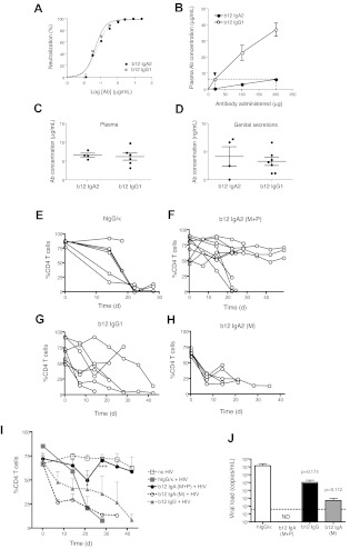
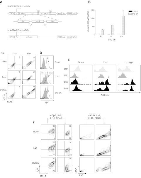
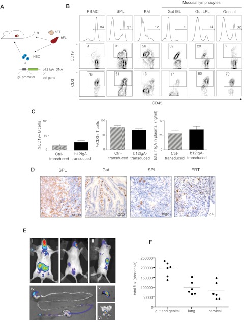
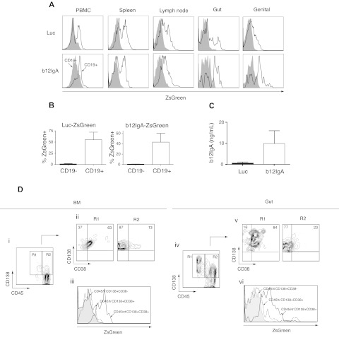
HIV-1 infections are generally initiated at mucosal sites. Thus, IgA antibody, which plays pivotal roles in mucosal immunity, might efficiently prevent HIV infection. However, mounting a highly effective HIV-specific mucosal IgA response by conventional immunization has been challenging and the potency of HIV-specific IgA against infection needs to be addressed in vivo. Here we show that the polymeric IgA form of anti-HIV antibody inhibits HIV mucosal transmission more effectively than the monomeric IgA or IgG1 form in a comparable range of concentrations in humanized mice. To deliver anti-HIV IgA in a continual manner, we devised a hematopoietic stem/progenitor cell (HSPC)-based genetic approach using an IgA gene. We transplanted human HSPCs transduced with a lentiviral construct encoding a class-switched anti-HIV IgA (b12-IgA) into the humanized bone marrow-liver-thymus (BLT) mice. The transgene was expressed specifically in B cells and plasma cells in lymphoid organs and mucosal sites. After vaginal HIV-1 challenge, mucosal CD4(+) T cells in the b12-IgA-producing mice were protected from virus-mediated depletion. Similar results were also obtained in a second humanized model, "human immune system mice." Our study demonstrates the potential of anti-HIV IgA in immunoprophylaxis in vivo, emphasizing the importance of the mucosal IgA response in defense against HIV/AIDS.


Figures

Figure 1
Figure 1
HIV-1 infection in humanized mice passively infused with b12-IgA or b12-IgG isotype. (A) Neutralization activity of anti-HIV human mAb b12-IgA. Reporter cell line TZM-bl cells that express CD4, CXCR4, CCR5, and a Tat-responsive reporter gene for luciferase were infected with 200 TCID50 of replication-defective pseudovirus containing Env (SF162.LS) in the presence of various concentrations of anti-HIV monoclonal antibodies. Neutralization activity was measured by the reduction in luciferase reporter gene expression after a single round of pseudovirus infection in TZM-bl cells in triplicate. b12-IgG1 and the recombinant b12-IgA2 were compared at indicated concentrations. (B) Antibody level in circulation after passive transfer. NSG-hu mice were injected intravenously with various concentrations of purified b12-IgA2 (pIgA: mIgA = 1:1, mass ratio) or b12-IgG1 and the blood was collected after 4 hours. The plasma antibody concentrations were measured using ELISA. (n = 4-6, mean ± SEM). (C-D) Concentrations of b12 antibodies in plasma (C) or genital secretions (D) at the time of challenge. NSG-hu mice were injected intravenously with either 200 μg of b12-IgA2 or 20 μg of b12-IgG1 per mouse. Blood and genital secretions were collected after 4 hours. (E-I) Peripheral CD4+ T cell loss after HIV-1 challenge in NSG-hu mice injected with different b12 antibody isotypes; (E) purified human IgG/κ control antibody (hIgG/κ); (F) b12-IgA2 includes both monomeric and polymeric IgA as described in (B), (b12IgA2 [M+P]); (G) b12-IgG1; (H) b12-IgA2 monomer only (b12IgA2 [M]). Mice were challenged intravaginally with HIV-1JR-CSF. (I) Average percent CD4+ T cells in CD3 T cells in PBMCs at each time points (n = 5-9). Statistical analysis was performed using unpaired t test. (*P = .0313 and ***P < .0001 between IgA [M+P] +HIV and control Ab [hIgG/κ] + HIV). (J) Plasma viral loads in HIV infected NSG-hu mice. HIV viral RNA was measured by Abbott HIV viral load test. Dotted line indicates the detection limit. (N/D indicates not detected; n = 4, indicated P values are from unpaired t test between Ab-treated groups with HIV and hIgG/κ-treated groups with HIV.)
Figure 2
Figure 2
Development of human B cells from HSPCs transduced with b12-IgA gene in vitro. (A) Schematic representation of the lentiviral constructs. Shown are pHAGE6-EEK-b12a-ZsGr and control vector pHAGE6-EEK-luc-ZsGr. The HIV-1 5′ LTR, b12 mAb heavy chain variable region (b12VH), human IgA2 heavy chain constant region α1, α2, α3 (CHα1, CHα2, CHα3), picornavirus-derived self-cleaving 2A peptide sequences (F2A and T2A), b12 κ light chain variable and constant region (b12VL-CLκ), human immunoglobulin J chain (J), the woodchuck hepatitis virus posttranscriptional regulatory element (WPRE) and 3′ LTR are indicated. For control vectors, the b12-IgA sequence was replaced by the luciferase gene in the same lentiviral vector. The human κ light chain promoter preceded by enhancers and matrix association regions is denoted as iEκ. The vector, pHAGE6-EEK-b12a-ZsGr contains a b12-IgA tri-cistronic cassette and a reporter gene ZsGreen (ZsGr) linked via IRES for simultaneous expression. (B) Secretion of human monoclonal antibody b12-IgA in human cells. Secreted human IgA in culture supernatant from 293T cells, which were transfected with either the lentiviral vector carrying b12-IgA or control vector (FUGW), were measured by human IgA ELISA at multiple time points. (C) CD10+CD19+ pro–B-cell development from HSPCs. Lentiviral transduction and in vitro human B lymphopoiesis culture were performed as previously described. CD34+ HSPCs transduced with either pHAGE6-EEK-luc-ZsGr (luc) or pHAGE6-EEK-b12a-ZsGr (b12-IgA) lentiviral constructs or left uninfected (none) cells were primed with IL-3, Flt3 ligand, thrombopoietin, SCF, and G-CSF for 5 days and then cocultured with MS5 stromal cells for indicated time points (C-E). Contour plots show CD10CD19 cell population at 14 days (left, D14) and emerging CD10+CD19+ pro–B-cell population after 21 days (right, D21) of culture. (D) Increased surface IgM expression. Data shown are gated on CD19+ cells from day 14 (D14) or day 32 (D32) culture. (E) Transgene expression during B-cell lymphopoiesis. Transferred luciferase or b12-IgA gene expression was assessed with the fluorescence of coexpressed ZsGreen protein using flow cytometry at indicated time points; day 14 (D14), day 21 (D21), day 32 (D32), and day 49 (D49) of HSPCs culture in the presence of MS5 cells. (F) Development to B-cell blast from mature B cells by stimulation. After being cocultured with MS5 cells for 8 to 9 weeks, CD19+ B cells were isolated and stimulated as previously described. The 2 left panels of the contour plot show levels of IgM on CD19+ B cells before and after stimulation. The 2 right panels depict forward versus side scatter contour plots before and after stimulation. Data shown are gated on CD19+ cells.
Figure 3
Figure 3
Generation of hu-BLT mice expressing b12-IgA through HSPC gene transfer. (A) A schematic diagram of the generation of hu-BLT mice transduced with human b12-IgA gene (hu-BLT-b12a mice). (B) Peripheral and mucosal reconstitution of a human immune system in hu-BLT-b12a mice. Flow cytometry shows human cell engraftment in various tissues isolated at 14 to 16 weeks after transplantation; PBMC indicates peripheral blood mononuclear cells; SPL, spleen; BM, bone marrow; Gut IEL, intestinal intraepithelial lymphocytes; Gut LPL, intestinal lamina propria lymphocytes; and genital, genital tract lymphocytes. (C) Percent engraftment of human B and T lymphocytes in periphery (Ctrl-transduced mice n = 7, b12-IgA-transduced mice n = 21) and the secretion of human IgA in the plasma (Ctrl-transduced mice, n = 5, b12-IgA-transduced mice, n = 12) of hu-BLT mice either transduced with b12-IgA gene or the control gene (mean ± SEM). (D) Immunohistochemical staining for human CD3 in spleen and small intestine tissues and human IgA-Producing cells in spleen and female reproductive tract (FRT) of hu-BLT-b12a mice. Samples were examined on an Olympus BX-51 microscope (20× objective lens) and photographed using a Spot Digital Camera. (E) Bioluminescence images of hu-BLT mice transduced with IgL chain promoter-driven luciferase transgene. The representative images of live animals displayed the distribution of human B-lymphocytes derived from transplanted human HSPCs that express transgenes; (Ei) ventral view of reference, (Eii) dorsal view of reference, and (Eiii) lateral view of reference. Strong bioluminescent signals from mucosal associated lymphoid tissues near gut, lung and genital tract area are seen in ventral view (Ei). Secondary lymphoid tissue (spleen) signals are seen in dorsal (Eii) and lateral view (Eiii). A luciferin-injected mouse was sacrificed after 5 minutes of incubation and dissected immediately to excise organs and tissues. The representative images of GI tract (Eiv), spleen (Ev), and genital tract (Evi) show tissue specificity of transgene expression. (F) Luminescent signals from hu-BLT mice described above at 18 weeks after transplantation. Signals recorded in photons/second were acquired from each indicated areas in the whole animal image.
Figure 4
Figure 4
b12-IgA transgene expression in B cells and plasma cells in mucosal and lymphoid tissues. (A) Human B cell (CD45+CD19+)–specific expression of transgene in hu-BLT mice. Gut-homing lymphocytes were isolated from the epithelia and lamina propria of the small and large intestine and genital tract-homing lymphocytes were isolated from the entire genital tract except the testis or ovary. Flow cytometry of the ZsGreen reporter gene expression of the lymphocytes isolated from PBMC, spleen, lymph node, gut and genital tract. The ZsGreen fluorescence of CD19 cells (filled histogram) and CD19+ (solid lines) was analyzed after gating on CD45+ human leukocytes. (B) Percent transgene expression of CD19 (black) and CD19+ (white) cells from peripheral blood of hu-BLT mice transduced with Luc-ZsGreen vector or b12-IgA-ZsGreen vector (n = 4-5, mean ± SEM). (C) Enzyme-linked immunosorbent assay of the secreted b12-IgA in plasma of hu-BLT mice transduced with Luc-ZsGreen vector (black) or b12-IgA-ZsGreen vector (white) at 10 weeks posttransplantation of human HSPCs (n = 6-8, mean ± SEM). (D) Plasma cell development from transplanted human HSPCs in hu-BLT mice transduced with b12-IgA-ZsGreen and transgene expression in long-lived plasma cells. Flow cytometry of bone marrow mononuclear cells (Di-iii) and gut lymphocytes (Div-Dvi) isolated from 22 to 24 week posttransplanted hu-BLT mice, with gating on CD45+ cells. The contour plots (Di,iv) show CD138+CD45int plasma cells (R1) and CD138+CD45hi (R2) plasma cells. R1 and R2 cell populations were then separately plotted by CD38 and CD138 expression (Dii,v). Histograms (Diii,vi) display reporter gene ZsGreen expression in CD45hiCD138+CD38 cells, CD45hiCD138+CD38+ early plasma cells and CD45intCD138+CD38+ mature plasma cells.
Figure 5
Figure 5
Prevention of mucosal transmission of HIV-1 in hu-BLT mice transduced with b12-IgA. Humanized-BLT mice were challenged with R5 tropic HIV-1 (JR-CSF) through intravaginal route at 14 to 20 weeks after transplantation or left unchallenged (no virus). Peripheral blood of the mice was collected periodically. Mice were killed after 8 to 10 weeks of HIV-1 challenge and lymphocytes from various tissues were isolated and analyzed by flow cytometry. (A) Protection of mucosal CD4+ T cells in hu-BLT-b12a mice from HIV-1 infection. Flow cytometry of mucosal lymphocytes isolated from intestinal intraepithelium (gut IEL), intestinal lamina propria (gut LPL) and female genital tract (genital) of hu-BLT mice that are transduced with b12-IgA gene (BLT-b12a) or with control gene (BLT-Ctrl). (B) Proportion of CD4+ T cells of primary (BM indicates bone marrow) or secondary (SPL indicates spleen) lymphoid organs and periphery. Cells were pregated on CD45+CD3+ cells (A-B). (C) Percent CD4+ T cells in CD45+CD3+ human T cells in the tissues of hu-BLT mice after HIV-1 infection. Data are mean ± SEM (n = 3-5, *P ≤ .05, **P ≤ .01). (D) Changes of CD4/CD8 ratio in PBMCs of hu-BLT mice after HIV-1 mucosal challenge. Data are mean ± SEM at each time points (weeks after challenge) open circle: no challenge; purple circle: HIV-1 challenge in control gene-transduced hu-BLT mice; green circle: HIV-1 challenge in b12-IgA-transduced hu-BLT mice. Unpaired t test showed the b12-IgA transduced group had significantly higher CD4:CD8 ratios than the control vector transduced group after HIV challenge. (n = 3-6, P < .05, P = .0148) All data points in each group regardless of time were included in the t test. (E) Differential expression of CCR5, the HIV-1 coreceptor level in human CD4+ T cells in hu-BLT mice. Histograms show the proportion of CCR5+CD4+ T cells in PBMC, gut lymphocyte, genital tract lymphocytes of hu-BLT mouse model. (F) Immunohistochemical staining of HIV-1 p24 protein in indicated tissues of hu-BLT mice after HIV-1 mucosal challenge. (SPL: spleen, SI: small intestine) Samples were examined on an Olympus BX-51 microscope (40× objective lens) and photographed using a Spot Digital Camera. (G) P24+ cells were determined by counting immunohistochemically stained cells from genital and intestinal tract tissue sections of hu-BLT mice. (n = 3-5, mean ± SEM).

Similar articles

Cited by

References

    1. Cerutti A, Rescigno M. The biology of intestinal immunoglobulin A responses. Immunity. 2008;28(6):740–750. - PMC - PubMed
    1. Wolbank S, Kunert R, Stiegler G, Katinger H. Characterization of human class-switched polymeric (immunoglobulin M [IgM] and IgA) anti-human immunodeficiency virus type 1 antibodies 2F5 and 2G12. J Virol. 2003;77(7):4095–4103. - PMC - PubMed
    1. Wright A, Lamm ME, Huang YT. Excretion of human immunodeficiency virus type 1 through polarized epithelium by immunoglobulin A. J Virol. 2008;82(23):11526–11535. - PMC - PubMed
    1. Kirchhoff F. Immune evasion and counteraction of restriction factors by HIV-1 and other primate lentiviruses. Cell Host Microbe. 2010;8(1):55–67. - PubMed
    1. Xu W, Santini PA, Sullivan JS, et al. HIV-1 evades virus-specific IgG2 and IgA responses by targeting systemic and intestinal B cells via long-range intercellular conduits. Nat Immunol. 2009;10(9):1008–1017. - PMC - PubMed

Publication types

MeSH terms