. 2013 Mar;6(2):342-57.
doi: 10.1242/dmm.010140. Epub 2012 Oct 12.

Exacerbated neuronal ceroid lipofuscinosis phenotype in Cln1/5 double-knockout mice

Affiliations

Exacerbated neuronal ceroid lipofuscinosis phenotype in Cln1/5 double-knockout mice

Tea Blom et al. Dis Model Mech. 2013 Mar.

Abstract

Both CLN1 and CLN5 deficiencies lead to severe neurodegenerative diseases of childhood, known as neuronal ceroid lipofuscinoses (NCLs). The broadly similar phenotypes of NCL mouse models, and the potential for interactions between NCL proteins, raise the possibility of shared or convergent disease mechanisms. To begin addressing these issues, we have developed a new mouse model lacking both Cln1 and Cln5 genes. These double-knockout (Cln1/5 dko) mice were fertile, showing a slight decrease in expected Mendelian breeding ratios, as well as impaired embryoid body formation by induced pluripotent stem cells derived from Cln1/5 dko fibroblasts. Typical disease manifestations of the NCLs, i.e. seizures and motor dysfunction, were detected at the age of 3 months, earlier than in either single knockout mouse. Pathological analyses revealed a similar exacerbation and earlier onset of disease in Cln1/5 dko mice, which exhibited a pronounced accumulation of autofluorescent storage material. Cortical demyelination and more pronounced glial activation in cortical and thalamic regions was followed by cortical neuron loss. Alterations in lipid metabolism in Cln1/5 dko showed a specific increase in plasma phospholipid transfer protein (PLTP) activity. Finally, gene expression profiling of Cln1/5 dko cortex revealed defects in myelination and immune response pathways, with a prominent downregulation of α-synuclein in Cln1/5 dko mouse brains. The simultaneous loss of both Cln1 and Cln5 genes might enhance the typical pathological phenotypes of these mice by disrupting or downregulating shared or convergent pathogenic pathways, which could potentially include interactions of CLN1 and CLN5.


Figures

Fig. 1.
Fig. 1.
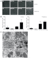
Impaired EB formation and differentiation of Cln1/5 dko iPS cells. (A) Microscopic images reveal the smaller size and more irregular shape of Cln1/5 dko EBs compared with wild-type EBs on day seven. (B) Statistical analysis of EB diameters shows significant difference in the EB size of wild-type and Cln1/5 dko clones. Columns present average EB size (n≥40); error bars represent s.e.m. *P<0.05, ***P<0.001. (C) EBs from 7-day-old mice were plated on gelatin-coated plates for spontaneous differentiation. Images show slower cellular outgrowth from Cln1/5 dko EBs compared with wild-type EBs, indicating delayed spontaneous differentiation potential of Cln1/5 dko EBs. Images were taken 3 days after plating. Scale bars: 500 μm.
Fig. 2.
Fig. 2.
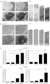
Accumulation of autofluorescent storage material in cerebral cortex and thalamus. (A) Confocal images of tissue sections from the cortex and thalamus show markedly increased autofluorescence in 3-month-old Cln1/5 dko mice. Scale bar: 100 μm. (B) Thresholding image analysis revealed higher levels, although not significantly, of autofluorescent storage material in the cortex and thalamus of Cln1/5 dko mice. (C) Electron micrographs of the storage material in the Cln1/5 dko, Cln1 ko and Cln5 ko brains. (C1) Cortical neuron of 3-month-old Cln1/5 dko mouse shows numerous storage bodies (indicated by arrows) and mitochondria in the perinuclear area. (C2) At higher magnification, ultrastructure of the neuronal storage material is clearly visible, showing typical granular osmiophilic deposits, GRODs, with tiny grains inside. The storage material is surrounded by a double membrane. (C3) Within the thalamus of 3-month-old Cln1/5 dko mice, some neurons were loaded with storage bodies, which were more electron-dense than those in the cerebral cortex. (C4) Higher magnification of the thalamic storage bodies shows typical GRODs surrounded by a double membrane. (C5) Ultrastructure of the storage material within cortical neurons of 3-month-old Cln1 ko mice closely resembles the GRODs seen in the cerebral cortex of Cln1/5 dko mice, although the storage deposits are less abundant in Cln1 ko mice than in Cln1/5 dko mice at this age. (C6) In the cortex of 3-month-old Cln5 ko mice, the neuronal storage material is composed of membranous profiles with rectilinear and fingerprint-like features. Magnification 10,000× in C1 and C3; 40,000× in C2 and C4-6. Scale bars: 2 μm (C1,C3); 1 μm (C2,C4-C6). s, storage bodies; m, mitochondria; N, nucleus.
Fig. 3.
Fig. 3.
Increased astrocytosis and widespread microglial activation in the thalamocortical system of 3-month-old Cln1/5 dko mice. (A) Immunohistochemical staining for the astrocytic marker GFAP in 3-month-old wild-type, Cln5 ko, Cln1 ko and Cln1/5 dko mice. Representative images from VPM/VPL and S1BF show pronounced and widespread astrocytosis in Cln1 ko and Cln1/5 dko brains, and elevated GFAP expression in the Cln5 ko brain. Insets from Cln5 ko, Cln1 ko and Cln1/5 dko show the morphology of GFAP-positive hypertrophic astrocytes with enlarged soma and thickened processes. (B) Immunohistochemical staining for the microglial marker CD68 in 3-month-old wild-type, Cln5 ko, Cln1 ko and Cln1/5 dko mice. Widespread activation of microglia in VPM/VPL and S1BF was evident in all three mouse models, being most elevated in Cln1 ko and Cln1/5 dko brains. Insets from Cln5 ko, Cln1 ko and Cln1/5 dko show the morphology of activated CD68-positive microglia, with swollen soma and shortened processes. Scale bars: 50 μm (insets) or as indicated. (C) Quantification of increased microglial activation (CD68 immunoreactivity) and (D) astrocytosis (GFAP immunoreactivity) in 3-month-old Cln1/5 dko mice. Thresholding image analysis confirms the significantly increased GFAP expression in the thalamic VPM/VPL and cortical S1BF regions of Cln1/5 dko mouse brain, compared with wild-type controls. CD68 expression was significantly increased in the S1BF of Cln1/5 dko mouse brain compared with wild-type brain. Error bars represent s.e.m. **P<0.01, *** P<0.001.
Fig. 4.
Fig. 4.
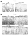
Defective myelination in the cortical laminae of Cln5 ko, Cln1 ko and Cln1/5 dko mouse brain. (A) Immunohistochemical staining with MBP revealed loss of MBP immunopositivity in Cln1/5 dko cortex at the age of 1 month, especially in the most superficial laminae of S1BF (laminae II and III). (B) All mouse models showed less MBP immunopositivity in the superficial laminae of S1BF than wild-type mice at the age of 3 months. Scale bar: 100 μm.
Fig. 5.
Fig. 5.
Early loss of cortical neurons in Cln1/5 dko mice. Unbiased optical fractionator estimates of the number of Nissl-stained neurons of 3-month-old mice in S1BF lamina IV (A), lamina V (B), lamina VI (C) and VPM/VPL (D) revealed significant loss of neurons in S1BF lamina VI of Cln1/5 dko brain. No significant loss of cortical S1BF neurons in laminae IV and V, or thalamic VPL/VPL neurons, was observed. *P<0.05.
Fig. 6.
Fig. 6.
Lipid profile analysis from 1-month-old wild-type, Cln1 ko, Cln5 ko and Cln1/5 dko mouse plasma. Cholesterol (Chol), phospholipid (PL), triglyceride (Trig) and apoA-1 levels (A) and PLTP activity (B) were determined from individual wild-type, Cln1 ko, Cln5 ko and Cln1/5 dko mouse plasma samples. PL levels, as well as PLTP activity, were significantly increased in Cln1/5 dko mice. Values plotted as mean + s.e.m. *P<0.05, **P<0.01, ***P<0.001.
Fig. 7.
Fig. 7.
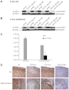
Westernblot analysis of α-synuclein levels in 3-month-old wild-type and Cln1/5 dko mouse brain. (A) In the cortex, α-synuclein protein expression was completely absent in Cln1/5 dko brains compared with wild-type brains. (B) α-Synuclein expression in cerebral lysates was absent in three out of five Cln1/5 dko samples. (C) Quantification of α-synuclein protein expression. Densitometric analysis shows a significant decrease in the expression of α-synuclein in the Cln1/5 dko cortex and cerebrum compared with the wild-type brains. (D) Immunohistochemical analysis of α-synuclein in 3-month-old wild-type, Cln1 ko, Cln5 ko and Cln1/5 dko brains. There was a marked reduction in α-synuclein staining in the neuropil of Cln1/5 dko cerebral cortex. Hippocampal α-synuclein staining was also decreased in the Cln1/5 dko brain. In addition, slightly reduced α-synuclein staining was observed in the cortical neuropil and hippocampus of the Cln5 ko brain. Scale bar: 50 μm. *P<0.05, ***P<0.001.

Similar articles

Cited by

References

    1. Abeliovich A., Schmitz Y., Fariñas I., Choi-Lundberg D., Ho W. H., Castillo P. E., Shinsky N., Verdugo J. M., Armanini M., Ryan A., et al. (2000). Mice lacking alpha-synuclein display functional deficits in the nigrostriatal dopamine system. Neuron 25, 239-252 - PubMed
    1. Ahtiainen L., Kolikova J., Mutka A. L., Luiro K., Gentile M., Ikonen E., Khiroug L., Jalanko A., Kopra O. (2007). Palmitoyl protein thioesterase 1 (Ppt1)-deficient mouse neurons show alterations in cholesterol metabolism and calcium homeostasis prior to synaptic dysfunction. Neurobiol. Dis. 28, 52-64 - PubMed
    1. Bible E., Gupta P., Hofmann S. L., Cooper J. D. (2004). Regional and cellular neuropathology in the palmitoyl protein thioesterase-1 null mutant mouse model of infantile neuronal ceroid lipofuscinosis. Neurobiol. Dis. 16, 346-359 - PubMed
    1. Camp L. A., Hofmann S. L. (1993). Purification and properties of a palmitoyl-protein thioesterase that cleaves palmitate from H-Ras. J. Biol. Chem. 268, 22566-22574 - PubMed
    1. Chrast R., Saher G., Nave K. A., Verheijen M. H. (2011). Lipid metabolism in myelinating glial cells: lessons from human inherited disorders and mouse models. J. Lipid Res. 52, 419-434 - PMC - PubMed

Publication types

MeSH terms