

Klf10 inhibits IL-12p40 production in macrophage colony-stimulating factor-induced mouse bone marrow-derived macrophages
- PMID: 23065757
- PMCID: PMC3842096
- DOI: 10.1002/eji.201242697
Klf10 inhibits IL-12p40 production in macrophage colony-stimulating factor-induced mouse bone marrow-derived macrophages
Abstract
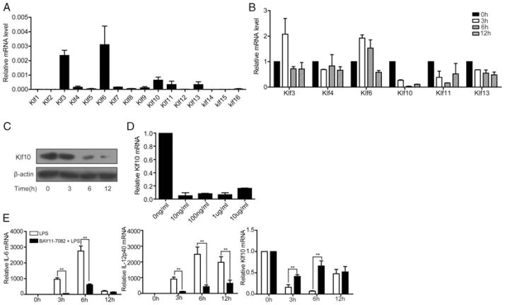
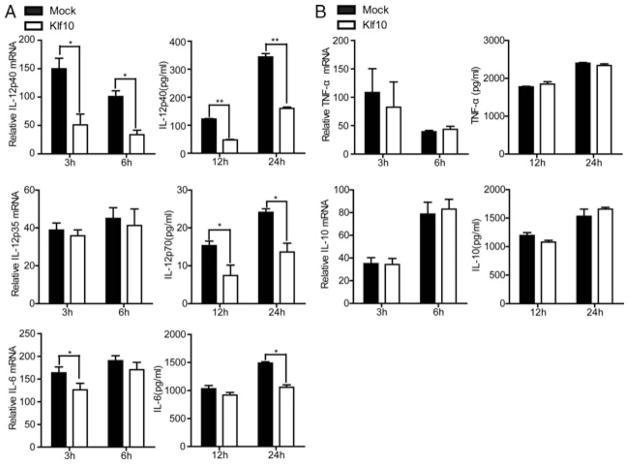
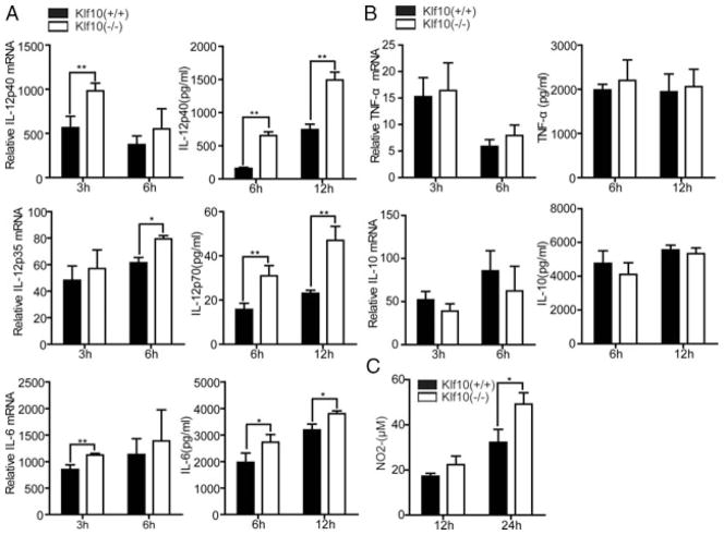
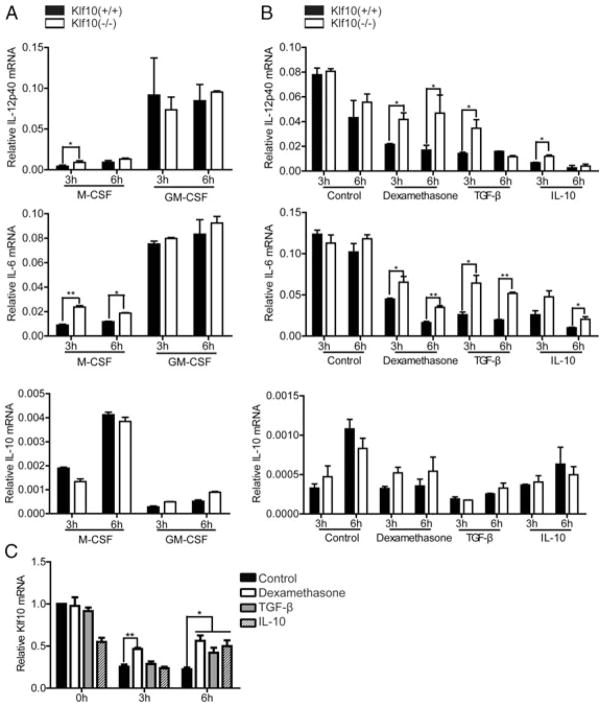
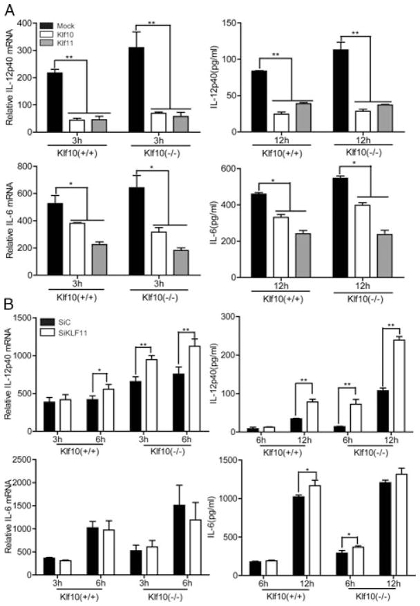
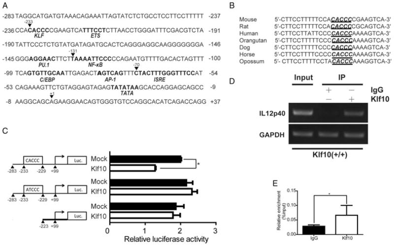
Bone marrow-derived macrophages (BMMs) treated with granulocyte-macrophage colony-stimulating factor (GM-CSF) or macrophage colony-stimulating factor (M-CSF), differentiate into GM-CSF-induced mouse bone marrow-derived macrophages (GM-BMMs) or M-CSF-induced mouse bone marrow-derived macrophages (M-BMMs), which have an M1 or M2 profile, respectively. GM-BMMs produce large amounts of proinflammatory cytokines and mediate resistance to pathogens, whereas M-BMMs produce antiinflammatory cytokines that contribute to tissue repair and remodeling. M-BMMs stimulated with lipopolysaccharide (LPS) are in an antiinflammatory state, with an IL-12(low) IL-10(high) phenotype. However, the regulation of this process remains unclear. Klf10 belongs to the family of Krüppel-like transcription factors and was initially described as a TGF-β inducible early gene 1. IL-12p40 is upregulated in LPS-stimulated M-BMMs from Klf10-deficient mice, but downregulated during Klf10 overexpression. Klf11, another member of the Krüppel-like factor family, can also repress the production of IL-12p40. Furthermore, Klf10 binds to the CACCC element of the IL-12p40 promoter and inhibits its transcription. We have therefore identified Klf10 as a transcription factor that regulates the expression of IL-12p40 in M-BMMs.
© 2012 WILEY-VCH Verlag GmbH & Co. KGaA, Weinheim.
Conflict of interest statement
Figures
Similar articles
-
Krüppel-like factor KLF10 deficiency predisposes to colitis through colonic macrophage dysregulation.Am J Physiol Gastrointest Liver Physiol. 2015 Dec 1;309(11):G900-9. doi: 10.1152/ajpgi.00309.2015. Epub 2015 Oct 15. Am J Physiol Gastrointest Liver Physiol. 2015. PMID: 26472224 Free PMC article.
-
Proteomic Analysis Reveals Distinct Metabolic Differences Between Granulocyte-Macrophage Colony Stimulating Factor (GM-CSF) and Macrophage Colony Stimulating Factor (M-CSF) Grown Macrophages Derived from Murine Bone Marrow Cells.Mol Cell Proteomics. 2015 Oct;14(10):2722-32. doi: 10.1074/mcp.M115.048744. Epub 2015 Jul 30. Mol Cell Proteomics. 2015. PMID: 26229149 Free PMC article.
-
Bone marrow-derived Kruppel-like factor 10 controls reendothelialization in response to arterial injury.Arterioscler Thromb Vasc Biol. 2013 Jul;33(7):1552-60. doi: 10.1161/ATVBAHA.112.300655. Epub 2013 May 16. Arterioscler Thromb Vasc Biol. 2013. PMID: 23685559 Free PMC article.
-
Klf10 and Klf11 as mediators of TGF-beta superfamily signaling.Cell Tissue Res. 2012 Jan;347(1):65-72. doi: 10.1007/s00441-011-1186-6. Epub 2011 May 17. Cell Tissue Res. 2012. PMID: 21574058 Review.
-
Functional role of KLF10 in multiple disease processes.Biofactors. 2010 Jan-Feb;36(1):8-18. doi: 10.1002/biof.67. Biofactors. 2010. PMID: 20087894 Free PMC article. Review.
Cited by
-
RNA-Seq transcriptomic profiling of primary murine microglia treated with LPS or LPS + IFNγ.Sci Rep. 2018 Oct 31;8(1):16096. doi: 10.1038/s41598-018-34412-9. Sci Rep. 2018. PMID: 30382133 Free PMC article.
-
KLF feedback loops in innate immunity.Front Immunol. 2025 Jun 4;16:1606277. doi: 10.3389/fimmu.2025.1606277. eCollection 2025. Front Immunol. 2025. PMID: 40534851 Free PMC article. Review.
-
Genetic deletion of Krüppel-like factor 11 aggravates traumatic brain injury.J Neuroinflammation. 2022 Nov 19;19(1):281. doi: 10.1186/s12974-022-02638-0. J Neuroinflammation. 2022. PMID: 36403074 Free PMC article.
-
SUMOylation of sPRDM16 promotes the progression of acute myeloid leukemia.BMC Cancer. 2015 Nov 11;15:893. doi: 10.1186/s12885-015-1844-2. BMC Cancer. 2015. PMID: 26559765 Free PMC article.
-
Cxxc Finger Protein 1 Positively Regulates GM-CSF-Derived Macrophage Phagocytosis Through Csf2rα-Mediated Signaling.Front Immunol. 2018 Aug 14;9:1885. doi: 10.3389/fimmu.2018.01885. eCollection 2018. Front Immunol. 2018. PMID: 30154795 Free PMC article.
References
-
- Martinez FO, Sica A, Mantovani A, Locati M. Macrophage activation and polarization. Front Biosci. 2008;13:453–461. - PubMed
-
- Gordon S. Alternative activation of macrophages. Nat Rev Immunol. 2003;3:23–35. - PubMed
-
- Krausgruber T, Blazek K, Smallie T, Alzabin S, Lockstone H, Sahgal N, Hussell T, et al. IRF5 promotes inflammatory macrophage polarization and TH1-TH17 responses. Nat Immunol. 2011;12:231–238. - PubMed
Publication types
MeSH terms
Substances
Grants and funding
LinkOut - more resources
Full Text Sources
Molecular Biology Databases
Research Materials
