. 2012 Nov 15;189(10):4870-80.
doi: 10.4049/jimmunol.1201296. Epub 2012 Oct 12.

NK cell lytic granules are highly motile at the immunological synapse and require F-actin for post-degranulation persistence

Affiliations

NK cell lytic granules are highly motile at the immunological synapse and require F-actin for post-degranulation persistence

Emily M Mace et al. J Immunol. .

Abstract

The formation of a dynamic, actin-rich immunological synapse (IS) and the polarization of cytolytic granules toward target cells are essential to the cytotoxic function of NK cells. Following polarization, lytic granules navigate through the pervasive actin network at the IS to degranulate and secrete their toxic contents onto target cells. We examined lytic granule motility and persistence at the cell cortex of activated human NK cells, using high-resolution total internal reflection microscopy and highly quantitative analysis techniques. We illustrate that lytic granules are dynamic and observe substantial motility at the plane of the cell cortex prior to, but not after, degranulation. We also show that there is no significant change in granule motility in the presence of Latrunculin A (which induces actin depolymerization), when added after granule polarization, but that there is a significant decrease in lytic granule persistence subsequent to degranulation. Thus, we show that lytic granules are highly dynamic at the cytolytic human NK cell IS prior to degranulation and that the persistence of granules at the cortex following exocytosis requires the integrity of the synaptic actin network.


Figures
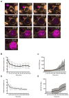
Figure 1
Figure 1. Lytic granule polarization precedes persistence at the NK immunological synapse prior to target cell death
YTS GFP-actin (A, B, C) or NK92 GFP-tubulin (D, E) NK cells were loaded with LysoTracker Red (red) and incubated with CellMask labeled 721.221 (A, B, C) or K562 (D, E) target cells in the presence of SYTOX Blue (blue) to detect cell death. Conjugates were imaged by confocal microscopy at 1 frame per minute for 90–130 minutes. A. Representative YTS GFP-actin-721.221 conjugate is shown at 10-minute intervals for 120 minutes. Scale bar=1 um. B. Time to granule polarization in YTS GFP-actin NK cells as measured by lytic granule centroid distance to IS (mean 41.5±12 minutes, n=10). C. Time to initiation of 721.221 target cell apoptosis as detected by SYTOX Blue entry is shown for 10 conjugates (mean 61.5 ± 10 minutes, n=10). D. Time to granule polarization in NK92 GFP-tubulin NK cells as measured by lytic granule centroid distance to IS (mean 24.2 minutes, n=6). E. Time to initiation of K562 target cell apoptosis as detected by SYTOX Blue entry is shown for 10 conjugates (mean 54.2 minutes, n=6). All representative images and analyses shown are from 4 independent experiments. Error bars indicate SD.
Figure 2
Figure 2
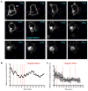
Visualization of NK cell lytic granule polarization and degranulation by live cell confocal microscopy. NK92 cells expressing pHlourin-LAMP1 (green) were loaded with LysoTracker Red (red) and incubated with Cell Mask labeled K562 cells as in Figure 1. Conjugates were imaged by confocal microscopy at 1 frame per minute for 90–130 minutes. A. One representative conjugate from 4 independent experiments is shown at 10-minute intervals for 60 minutes. One degranulation event is shown at 25 minutes. Scale bar = 1 μm. B. Granule polarization in representative NK92 pHlourin-LAMP1 cell shown in A as measured in Figure 1. Red vertical dashed lines indicate brief individual degranulation events prior to the sustained event beginning at 25 minutes (heavy red dashed line). C. Mean granule polarization from 4 independent experiments (n = 4) with vertical red lines indicating initial degranulation events from each conjugate analyzed. Error bars indicate SD.
Figure 3
Figure 3. Lytic granules navigate the cell cortex prior to degranulation
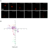
NK92 cells expressing pHlourin-LAMP1 (green) were loaded with LysoTracker Red (red) and activated by immobilized antibody to NKp30 and CD18. Cells were imaged by TIRFm at 6 frames per minute for 60–80 minutes. A. Representative NK92 lytic granule is shown at 5-minute intervals following 10 minutes of cell contact with the activating surface. Granules were tracked using Volocity software prior to and following degranulation as described in Materials and Methods. Pre-degranulation LysoTracker Red (red) and post-degranulation pHluorin-LAMP1 (green) tracks are shown in the final 55-minute image. Scale bar=1 μm. B. Overlay of LysoTracker Red tracks of 14 pre-degranulation events over 4 separate experiments. Lytic granule track from (A) shown in bold (red). C. Overlay of pHluorin-LAMP1 tracks of corresponding degranulation events. Lytic granule track from (A) shown in bold (green).
Figure 4
Figure 4. Synaptic lytic granules show greater motility prior to degranulation
Granule tracks were analyzed using Volocity software as described in Materials and Methods. Granule track length (A), track velocity (B), displacement (C), and displacement rate (D) from 14 events pre- (LysoTracker Red, left) and post-degranulation (pHlourin-LAMP1, right) are shown. Representative NK92 lytic granules from Figure 3A are indicated by open diamonds. Mean ± SD are shown. Differences between Lysotracker Red and pHlourin-LAMP1 granule tracks were significant (p <0.0001, two-tailed t test). Results are from 4 independent experiments.
Figure 5
Figure 5. Lytic granules that do not degranulate show normal synaptic motility
Lytic granules demonstrating no observed degranulation were analyzed. A. Representative NK92 lytic granule cropped from image sequence is shown at 5-minute intervals following 10 minutes of contact. Lysotracker Red (red) track denoting all observed timepoints is shown in final 55-minute image. Scale bar=1 μm. B. Overlay of LysoTracker Red tracks of 10 lytic granules over 4 separate experiments. Representative granule track from (A) is depicted in bold (red).
Figure 6
Figure 6. Characteristics of synaptic lytic granule motility that do not degranulate
Comparative measurements of LysoTracker Red motility in lytic granules for which degranulation or no degranulation was observed. Length (A), track velocity (B), displacement (C), displacement rate (D), and persistence time (E) plotted for 10 granules observing no degranulation (LysoTracker Red only). Data is shown alongside measurements obtained from degranulation events previously illustrated in Figure 4 (i.e., the same results in Figure 3 for degranulating granules). The representative NK92 granule from Figure 5A is indicated in LysoTracker Red data set for comparison to the other granules measured, but for which image sequences are not shown (open diamond). F) Velocities of granules that degranulate (black) or do not (red) were measured at one minute intervals. Mean ± SD are depicted. The means of the LysoTracker Red data sets were not significantly different (p>0.05, two-tailed t test). Results are shown from 4 independent experiments.
Figure 7
Figure 7. Effect of actin depolymerization upon synaptic lytic granule kinetics
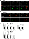
NK92 cells expressing pHlourin-LAMP1 (green) were loaded with LysoTracker Red (red) and activated upon immobilized antibody to NKp30 and CD18. Cells were imaged by TIRF microscopy at 6 frames per minute for 60–80 minutes. A representative NK92 lytic granule cropped from the image sequence is shown at 5-minute intervals following 5–10 minutes of contact-induced activation. LysoTracker Red and pHluorin-LAMP1 tracks depicting the course of the granule over all timepoints are shown in final 55-minute image. Scale bars=1 μm. A. Vehicle control (DMSO) was added 10–20 minutes following the addition of cells to the imaging chamber. The white circle indicates granules location in frames 1–5. B. LatA was added between 10 and 15 minutes for a final concentration of 10 μM. C. Measured mean characteristics of synaptic lytic granule motility before (black bars) and after (white bars) degranulation. Mean track length, track velocity, displacement, displacement rate, and timespan of lytic granules are all shown relative to the respective DMSO values, which have been normalized to 1. Mean ± SD are shown. Significant differences between DMSO- and Latrunculin A-treated granule tracks are marked with an asterisk (p<0.05, two-tailed t test). Results shown are from 4 independent experiments.
Figure 8
Figure 8. The synaptic actin network is required for the persistence of degranulation
A. The lifetime of lytic granules post-degranulation in NK cells treated with LatA or DMSO control. Time points reflect amount of time elapsed post-degranulation (marked by the appearance of LAMP1-phluorin) with vertical drops indicating disappearance of the granule from TIRF field. Vertical ticks indicate granules persisting to the end of the imaging sequence. B. Area of the observed lytic granules in cells treated with DMSO (solid black), or LatA (dashed red). C. Sum fluorescent intensity of lytic granules in NK92 cells expressing pHluorin-LAMP1. Cells were treated with DMSO (solid black), or LatA (dashed red) as per Figure 7. Note that sum fluorescent intensity is a function of both the area and mean fluorescent intensity of a lytic granule. Granule boundaries were defined using fluorescent intensity with 3 SD above background as a cutoff. Results shown are from 4 independent experiments.
Figure 9
Figure 9. Models for the role of F-actin in granule persistence and the varying behavior of lytic granules at the NK cell cortex
A) A LysoTracker Red-loaded LAMP1-pHlourin expressing granule is depicted approaching within cell cortex nearing a region within the F-actin network suitable for membrane access (as previously demonstrated (5, 7) (1). As docking and fusion occurs (2), F-actin acts as a tether to help anchor the granule at the membrane, although in both cases fusion results in the activation of LAMP1-phluorin and the subsequent appearance of green fluorescence. In addition, actin reorganization is likely to act in the generation of force to aid in the focused expulsion of granule contents (as supported by greater area*intensity of pHluorin-LAMP1 in control-compared to LatA-treated cells) (3) and the continued persistence of the degranulating granule at the cortex which we would propose is a feature of the interaction of the granule with the local F-actin network (4). B) A LysoTracker Red loaded LAMP1-pHlourin expressing granule approaches the cell membrane and docks with the aid of F-actin tethering (1, 2). This is followed by the approximation of the granule to the cell cortex, movement, then one of the outcomes depicted below. i) immediate exocytosis, ii) limited movement and exocytosis, iii) immediate withdrawal from the cortex or iv) limited movement before withdrawal.

Similar articles

Cited by

References

    1. Orange JS. Formation and function of the lytic NK-cell immunological synapse. Nat Rev Immunol. 2008;8:713–725. - PMC - PubMed
    1. Wulfing C, Purtic B, Klem J, Schatzle JD. Stepwise cytoskeletal polarization as a series of checkpoints in innate but not adaptive cytolytic killing. Proc Natl Acad Sci U S A. 2003;100:7767–7772. - PMC - PubMed
    1. Zarcone D, Prasthofer EF, Malavasi F, Pistoia V, LoBuglio AF, Grossi CE. Ultrastructural analysis of human natural killer cell activation. Blood. 1987;69:1725–1736. - PubMed
    1. Mentlik AN, Sanborn KB, Holzbaur EL, Orange JS. Rapid lytic granule convergence to the MTOC in natural killer cells is dependent on dynein but not cytolytic commitment. Mol Biol Cell. 2010;21:2241–2256. - PMC - PubMed
    1. Rak GD, Mace EM, Banerjee PP, Svitkina T, Orange JS. Natural killer cell lytic granule secretion occurs through a pervasive actin network at the immune synapse. PLoS Biol. 2011;9:e1001151. - PMC - PubMed

Publication types

MeSH terms