Review
. 2012 Jul-Aug;1(4):479-99.
doi: 10.1002/wdev.32.

Planar cell polarity signaling: coordination of cellular orientation across tissues

Affiliations
Review

Planar cell polarity signaling: coordination of cellular orientation across tissues

Jaskirat Singh et al. Wiley Interdiscip Rev Dev Biol. 2012 Jul-Aug.

Abstract

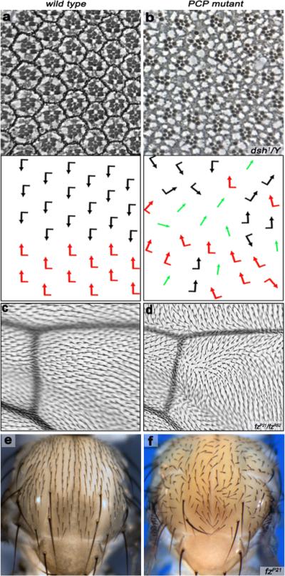
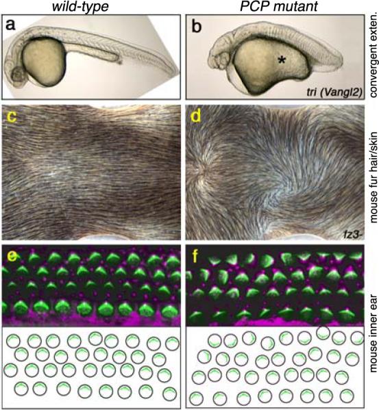
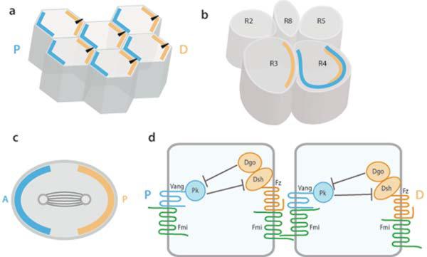
Establishment of Planar Cell Polarity (PCP) in epithelia, in the plane of an epithelium, is an important feature of the development and homeostasis of most organs. Studies in different model organisms have contributed a wealth of information regarding the mechanisms that govern PCP regulation. Genetic studies in Drosophila have identified two signaling systems, the Fz/PCP and Fat/Dachsous system, which are both required for PCP establishment in many different tissues in a largely non-redundant manner. Recent advances in vertebrate PCP studies have added novel factors of PCP regulation and also new cellular features requiring PCP-signaling input, including the positioning and orientation of the primary cilium of many epithelial cells. This review focuses mostly on several recent advances made in the Drosophila and vertebrate PCP field and integrates these within the existing PCP-signaling framework.


Figures

Figure 1
Figure 1. Examples of PCP in adult Drosophila tissues
(a–b) PCP features in the eye. Anterior is left and dorsal is up. Tangential eye sections showing wild-type adult eye (a) and a dsh1 eye (b) centered on the equator; bottom panels show schematic representations reflecting ommatidial orientation and polarity. Black and red arrows represent the dorsal and ventral chiral forms respectively while green arrows represent R3-R3 symmetrical clusters. In the PCP mutant (dsh in b) the arrangement of ommatidia is disorganized. (c–d) PCP aspects of wing patterning. Anterior is up and distal right. Each wing cell gives rise to an actin based hair (trichome) that is pointing distally in wild-type (c). Mutations in PCP genes (a fz example is shown in d) disrupt this near perfect orientation of wing hairs/trichomes, creating swirls and waves. (e–f) Aspects of PCP establishment on the thorax/notum. Anterior is up. Wild type adult thorax (e) showing mechano-sensory bristles, which are patterned uniformly across the notum and oriented in the anterior-posterior axis of the Drosophila body. In fz mutant (f) this regular pattern is randomized. In addition to the sensory bristles, all body wall cells form actin-rich trichomes (like in the wing), which are also oriented in the Anterior-posterior axis (not visible at this magnification).
Figure 2
Figure 2. Examples of PCP features in vertebrates
PCP features of convergent extension gastrulation movements in the zebrafish (a–b), the mouse skin (c–d) and the mouse inner ear (e–f). Anterior is right in all panels. Wild-type is on the left and PCP mutants in the right column. (a–b) Zebrafish embryos: the mutant PCP genotype is a maternal-zygotic mutant of trilobite/Vangl2. Note short and wide (fat) body axis in PCP mutant. The original pictures were provided by Brian Ciruna. (c–d) Dorsal view of mouse neck displaying the orientation of fur hair (and underlying skin) in wild-type (c) and mfz3 mutants (d). Note random waves and whorls in the fz3- genotype, compare to the normal anterior-posterior orientation in wild-type (c). (e–f) Orientation of sensory hair cells of the mouse choclea (inner ear). Each cell contains polarized bundles of actin-based stereocilia (green; labeled with phalloidin) and a tubulin based kinocilium (labeled with anti-acetylated tubulin; magenta). In PCP mutants these bundles still form but their orientation becomes randomized (f; Looptail/Vangl2 mutant). The lower panels show schematic representation of the cellular (actin bundle) orientation.. The original pictures of c–f were kindly provided by Jeremy Nathans.
Figure 3
Figure 3. Schematic Presentation of Asymmetric Core Fz/PCP Protein Localization
(a) Schematic of wing cells highlighting the epithelial nature of these cells. These hexagonal cells display PCP orientation in the proximal-distal axis). Single actin rich hairs (black arrowheads in each cell) project from the distal vertex of each cell, where the Fz-Dsh complex (orange) gets localized. The asymmetric localization of the core protein complexes at the end of PCP establishment (shown in blue and orange) serves as molecular markers for cell orientation. The blue complex contains Vang/Stbm, Pk and Fmi proteins, while the orange complex contains Fz, Dsh, Dgo, and Fmi (see panel d for molecular interactions). (b) Asymmetric distribution of core PCP proteins is also observed during eye patterning in the precursors to the R3 and R4 photoreceptors, which is a prerequisite for proper fate specification of R3 and R4, at the time of the 5-cell precluster posterior to the furrow (individual R-cells are numbered according to their final fate) (c) Example of a dividing sensory organ precursor (SOP) cell, showing polarized orientation of the mitotic spindle. The orientation of the spindle depends on the asymmetric localization of the core PCP proteins (shown in blue and orange as above). (d) Schematic presentation of asymmetric localization and molecular interactions of core PCP proteins across two wing epithelial cells. Fz–Dsh–Dgo–Fmi is enriched in a form of complex at the distal edges of cells, while the Vang–Pk–Fmi complex is concentrated to proximal edges of cells. Fz (orange) binds to Vang (blue) primarily via its CRD and this interaction is stabilized by homophilic interactions mediated by the atypical cadherin Fmi (green). Fz forms an intracellular complex with Dsh and Dgo, while Vang interacts intracellularly with Pk. Dsh and Dgo physically interact with each to promote Fz-Dsh signaling and can antagonize Pk. Diego competes with Prickle for binding to Dsh and thus antagonizes the inhibitory effect of Pk on Dsh.

References

    1. Gubb D, Garcia-Bellido A. A genetic analysis of the determination of cuticular polarity during development in Drosophila melanogaster. J Embryol Exp Morphol. 1982;68:37–57. - PubMed
    1. Lawrence PA, Shelton PM. The determination of polarity in the developing insect retina. J Embryol Exp Morphol. 1975;33:471–486. - PubMed
    1. Vinson CR, Adler PN. Directional non-cell autonomy and the transmission of polarity information by the frizzled gene of Drosophila. Nature. 1987;329:549–551. - PubMed
    1. Klein TJ, Mlodzik M. Planar cell polarization: an emerging model points in the right direction. Annu Rev Cell Dev Biol. 2005;21:155–176. - PubMed
    1. Lawrence PA, Struhl G, Casal J. Planar cell polarity: one or two pathways? Nat Rev Genet. 2007;8:555–563. - PMC - PubMed

Publication types

LinkOut - more resources