Review
. 2013 Jan;105(1):1-13.
doi: 10.1111/boc.201200044. Epub 2012 Dec 5.

Functional asymmetry in kinesin and dynein dimers

Affiliations
Review

Functional asymmetry in kinesin and dynein dimers

Katherine C Rank et al. Biol Cell. 2013 Jan.

Abstract

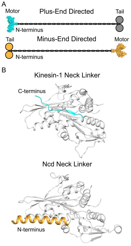
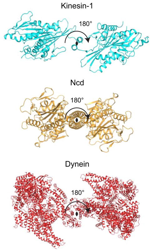
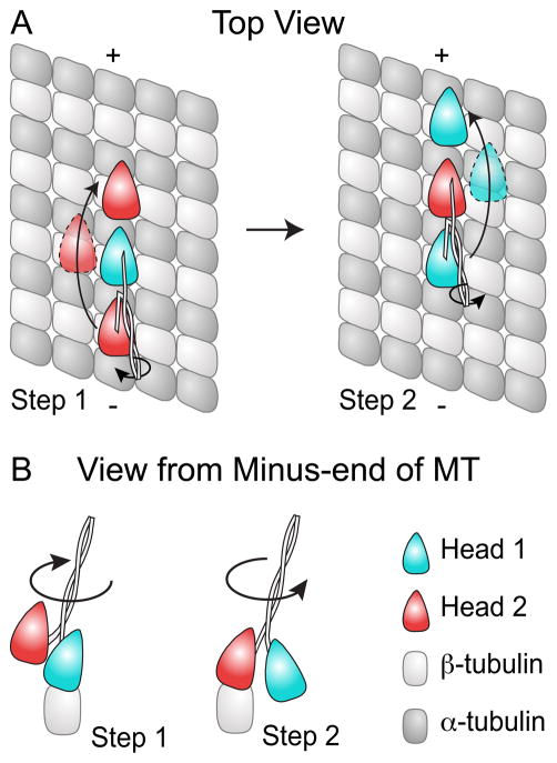
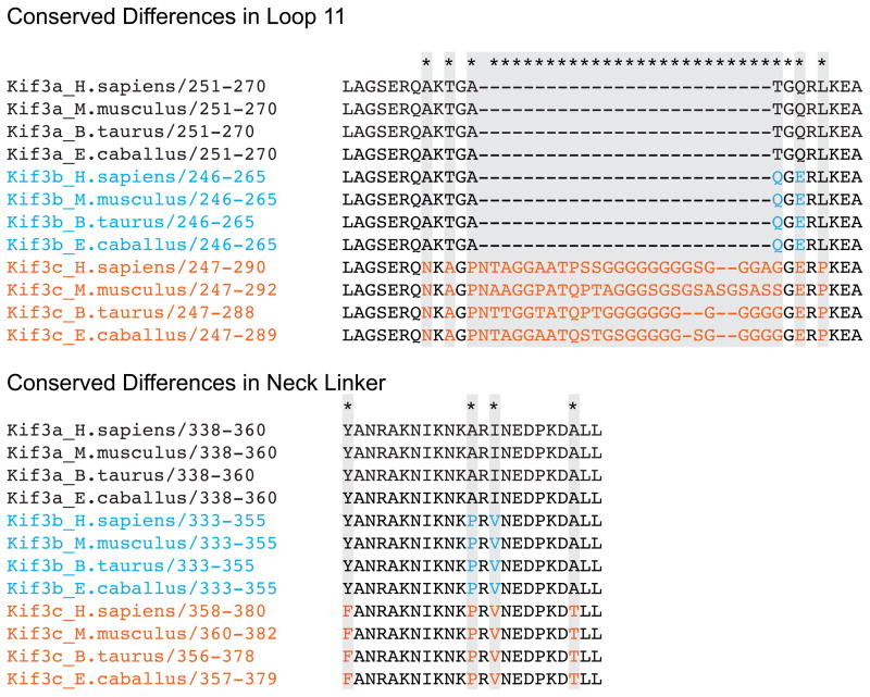
Active transport along the microtubule lattice is a complex process that involves both the Kinesin and Dynein superfamily of motors. Transportation requires sophisticated regulation much of which occurs through the motor's tail domain. However, a significant portion of this regulation also occurs through structural changes that arise in the motor and the microtubule upon binding. The most obvious structural change being the manifestation of asymmetry. To a first approximation in solution, kinesin dimers exhibit twofold symmetry, and microtubules exhibit helical symmetry. The higher symmetries of both the kinesin dimers and microtubule lattice are lost on formation of the kinesin-microtubule complex. Loss of symmetry has functional consequences such as an asymmetric hand-over-hand mechanism in plus-end-directed kinesins, asymmetric microtubule binding in the Kinesin-14 family, spatially biased stepping in dynein and cooperative binding of additional motors to the microtubule. This review focusses on how the consequences of asymmetry affect regulation of motor heads within a dimer, dimers within an ensemble of motors, and suggests how these asymmetries may affect regulation of active transport within the cell.


Figures

Figure 1
Figure 1. Topological Organization of Plus- and Minus-End Directed Kinesins
A. Overall topology of plus- and minus-end directed motors (cyan and orange, respectively) where plus-end directed motors contain N-terminal motor domains and C-terminal cargo domains, and minus-end directed motors contain N-terminal cargo domains and C-terminal motor domains. A surface representation of a dimeric Kinesin-1 motor domain (pdb accession 2Y65) is shown as a representation structure of a plus-end directed motor, and a surface representation of a dimeric Ncd motor domain (pdb accession 2NCD) is shown as a representative structure of a minus-end directed motor (Sablin et al., 1998; Kaan et al., 2011). The cargo domains of the plus-end directed motors are depicted in grey as some plus-end kinesins, such as Kinesin-1, utilize a second polypeptide chain for their cargo domains. B. Structure of the neck linker in Kinesin-1 and Ncd (pdb accession 2Y65 and 2NCD) show two radically different conformations. In plus-end directed motors, the neck linker is located C-terminal of the motor domain (grey) and exhibits a random coil and β-strand secondary structure. The depicted orientation represents a parallel docked position. In minus-end directed motors, the neck linker is located N-terminal to the motor domain (grey) and exists as an α-helix which forms a coiled-coil with the second head in the dimer. The depicted position represents a pre-powerstroke orientation. The neck linker region in minus-end directed motors is not as well defined as in plus-end directed motors; the region highlighted here represents the section of coiled-coil that makes direct contacts with the motor domain and is required for wild-type motility (Sablin et al., 1998; Rank et al., 2012).
Figure 2
Figure 2. Symmetry between Motor Domains in a Dimer
Cartoon representations of dimeric structures of Kinesin-1 (pdb accession 2Y5W, cyan), Ncd (2NCD, orange), and dynein (4AKG, red) viewed looking down the coiled-coil with the 2-fold axis depicted as a black oval (Sablin et al., 1998; Kaan et al., 2011; Schmidt et al., 2012). Both Kinesin-1 and dynein show close to a 2-fold symmetry between heads. Variation from 2-fold symmetry likely reflects flexibility within the neck linker/GST linker and effects of crystal packing. The Ncd structure shows crystallographic 2-fold symmetry where only one polypeptide is found within the asymmetric unit of the unit cell.
Figure 3
Figure 3. Motor Domain Trajectories during Asymmetric Hand-over-Hand Mechanism
(A) A schematic representation of the different trajectories experienced by kinesin dimer heads during an asymmetric hand-over-hand stepping motion. In this model, head 2 (red) travels to the left of the protofilament, when viewed from the minus-end of the microtubule, and a greater distance above the protofilament while stepping compared to head 1 (cyan), which swings to the right of the protofilament during its step. (B) Stepping of the heads also introduces twist within the coiled-coil; however as each step introduces twist in the opposite direction, the net rotation within the cycle is zero. This is more clearly illustrated in the view from the minus-end of the microtubule.
Figure 4
Figure 4. Sequence Conserved Differences within heterodimeric Kinesin-2 Motors
Kif3a, Kif3b, and Kif3c show conserved sequence differences between themselves. Conserved differences within the microtubule binding region, particularly loop 11, and the neck linker suggests that these motors have evolved to have different microtubule binding interactions and to respond differently to tension within the neck linker. It is possible that these differences within the neck linker optimize the motors to work at the different vertical loads, and that differences within the microtubule binding region bias one motor to bind first to the microtubule. Sequences aligned using T-coffee server (Notredame et al., 2000), and further analyzed utilizing Jalview (Clamp et al., 2004; Waterhouse et al., 2009).
Figure 5
Figure 5. A cryo-EM image of microtubules decorated with dimeric Ncd at subsaturating ratios reveals a cooperative binding mechanism in the axial direction, along individual protofilaments
While one of the microtubules is mostly decorated (I), partial decoration (III) and complete decoration (II) co-exist in the same area. A Fourier-filtered image of the microtubule and the motors in a side projection allows determination of the polarity of the microtubule according to the shape of the dimeric motor construct (Adapted from Figure 1 of (Wendt et al., 2002)).
Figure 6
Figure 6. Cooperative binding to the Microtubule
A. Kinesin-1 (cyan) dimers exhibit cooperative binding to the microtubule, and the presence of 16 nm layer lines in cryo-EM suggests this proceeds in an lateral manner (Vilfan et al., 2001). Kinesin-14 dimers (orange) also display cooperative binding; however in contrast to Kinesin-1, this appears to proceed along the protofilament (Wendt et al., 2002; Cope et al., 2010). The second head in the Kinesin-14 dimer is depicted as partially transparent since cooperative binding is only observed when one head of the dimer is bound; for Kar3Vik1, binding of both heads in the ADP state appeared to abolish cooperative binding (Rank et al., 2012). C. Kinesin-1 dimers also exhibit long range cooperative binding where binding of one dimer biases additional dimers to bind further towards the plus-end of the microtubule (Muto et al., 2005).

References

    1. Aizawa H, Sekine Y, Takemura R, Zhang Z, Nangaku M, Hirokawa N. Kinesin family in murine central nervous system. The Journal of cell biology. 1992;119:1287–1296. - PMC - PubMed
    1. Allingham JS, Sproul LR, Rayment I, Gilbert SP. Vik1 modulates microtubule-Kar3 interactions through a motor domain that lacks an active site. Cell. 2007;128:1161–1172. - PMC - PubMed
    1. Asbury CL, Fehr AN, Block SM. Kinesin moves by an asymmetric hand-overhand mechanism. Science. 2003;302:2130–2134. - PMC - PubMed
    1. Barrett JG, Manning BD, Snyder M. The Kar3p kinesin-related protein forms a novel heterodimeric structure with its associated protein Cik1p. Mol Biol Cell. 2000;11:2373–2385. - PMC - PubMed
    1. Carter AP, Cho C, Jin L, Vale RD. Crystal structure of the dynein motor domain. Science. 2011;331:1159–1165. - PMC - PubMed

Publication types

LinkOut - more resources