Review
. 2012 Dec;8(12):711-6.
doi: 10.1038/nrneurol.2012.210. Epub 2012 Oct 16.

The vascular neural network--a new paradigm in stroke pathophysiology

Affiliations
Review

The vascular neural network--a new paradigm in stroke pathophysiology

John H Zhang et al. Nat Rev Neurol. 2012 Dec.

Abstract

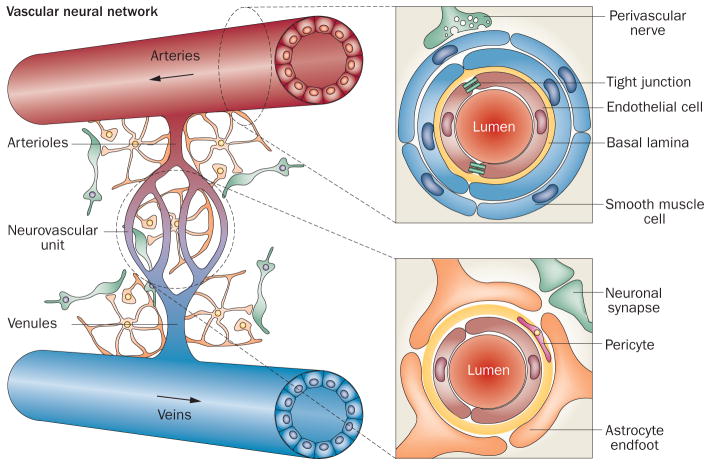
The concept of the neurovascular unit as the key brain component affected by stroke is controversial, because current definitions of this entity neglect mechanisms that control perfusion and reperfusion of arteries and arterioles upstream of the cerebral microcirculation. Indeed, although definitions vary, many researchers consider the neurovascular unit to be restricted to endothelial cells, neurons and glia within millimetres of the cerebral capillary microcirculation. This Perspectives article highlights the roles of vascular smooth muscle, endothelial cells and perivascular innervation of cerebral arteries in the initiation and progression of, and recovery from, ischaemic stroke. The concept of the vascular neural network-which includes cerebral arteries, arterioles, and downstream neuronal and glial cell types and structures-is introduced as the fundamental component affected by stroke pathophysiology. The authors also propose that the vascular neural network should be considered the main target for future therapeutic intervention after cerebrovascular insult.


Conflict of interest statement

Competing interests

The authors declare no competing interests.

Figures

Figure 1
Figure 1
The neurovascular unit as a component of the vascular neural network. The neurovascular unit consists of capillary endothelial cells, pericytes and basal lamina enveloped by astrocyte endfeet. Astrocytes communicate with adjacent neurons via metabolite exchange. The vascular neural network is larger than the neurovascular unit, as it also includes smooth muscle cells, noncapillary endothelial cells, perivascular nerves, fibroblasts, smooth muscle progenitor cells, and cells of the immune system—as well as collateral blood vessels, the rete vasorum, perivascular nerves and veins. The vascular neural network, therefore, comprises all cells and structures required to maintain cerebral blood flow under physiological and pathological conditions.
Figure 2
Figure 2
The evolving concept of the neurovascular unit. The concept of the neurovascular unit has changed considerably since its initial proposal as a triad of arteries, astrocytes and neurons in 1996. Vascular smooth muscle cells, endothelial cells and perivascular nerves were also included in the definition in some early publications. The definition jointly proposed by the NIH and National Institute of Neurological Disorders and Stroke in 2002 comprises endothelial cells, astrocytes and neurons, and subsequent publications highlighted physiological and pathophysiological roles for these cells within the neurovascular unit.*Indicates the initial 1996 publication. Indicates the 2002 NIH–National Institute of Neurological Disorders and Stroke publication.
Figure 3
Figure 3
Immunostained section of a cerebral artery showing the range of cell types present. The vessel lumen is lined with endothelial cells, and pericytes are visible in the subendothelial space. Vascular smooth muscle cells make up the medial layer, and perivascular nerves occur at the medial–adventitial border. The function and phenotype of these cell types are dynamically modulated by various factors released from brain parenchyma, fibroblasts and macrophages in the adventitia, perivascular nerves, vascular smooth muscle cells and endothelial cells (listed on the left). Abbreviations: IGF, insulin-like growth factor; BDNF, brain derived neurotrophic factor; FGF, fibroblast growth factor; NGF, nerve growth factor; NPY, neuropeptide Y; PDGF, platelet derived growth factor; TGF-β, transforming growth factor-β; VEGF, vascular endothelial growth factor.

References

    1. Lo EH, Dalkara T, Moskowitz MA. Mechanisms, challenges and opportunities in stroke. Nat Rev Neurosci. 2003;4:399–414. - PubMed
    1. Ginsberg MD. Neuroprotection for ischemic stroke: past, present and future. Neuropharmacology. 2008;55:363–389. - PMC - PubMed
    1. del Zoppo GJ. The neurovascular unit in the setting of stroke. J Intern Med. 2010;267:156–171. - PMC - PubMed
    1. Lo EH, Broderick JP, Moskowitz M. A tPA and proteolysis in the neurovascular unit. Stroke. 2004;35:354–356. - PubMed
    1. Iadecola C. Neurovascular regulation in the normal brain and in Alzheimer’s disease. Nat Rev Neurosci. 2004;5:347–360. - PubMed

Publication types