

Synthetic hydrogel scaffold is an effective vehicle for delivery of INFUSE (rhBMP2) to critical-sized calvaria bone defects in rats
- PMID: 23070779
- PMCID: PMC3565235
- DOI: 10.1002/jor.22243
Synthetic hydrogel scaffold is an effective vehicle for delivery of INFUSE (rhBMP2) to critical-sized calvaria bone defects in rats
Abstract
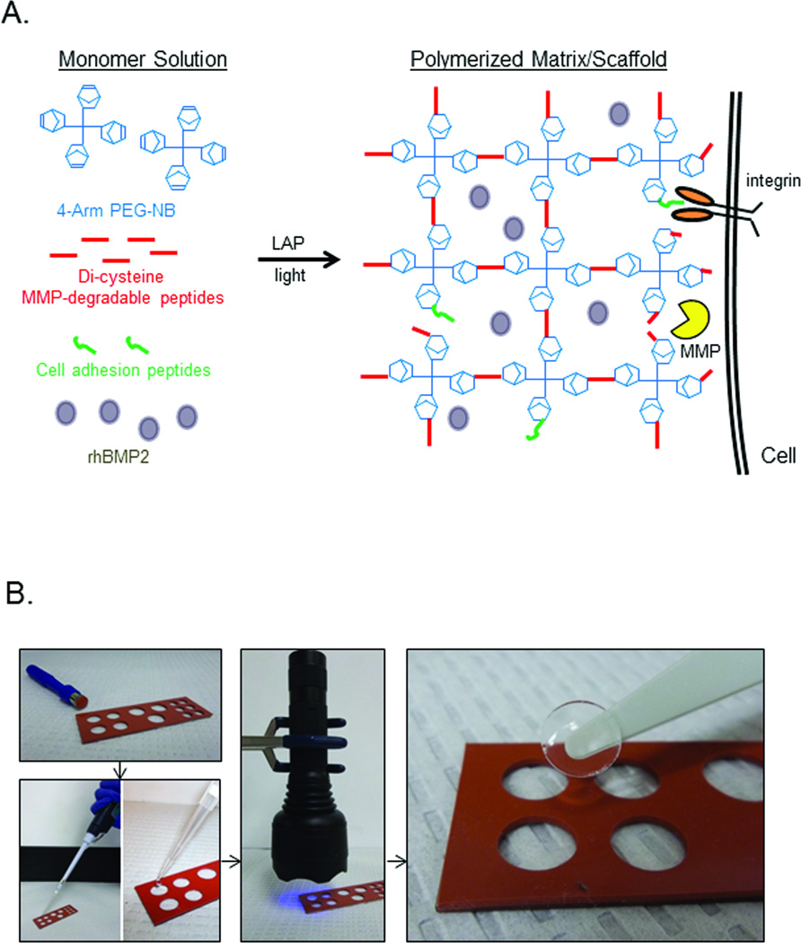
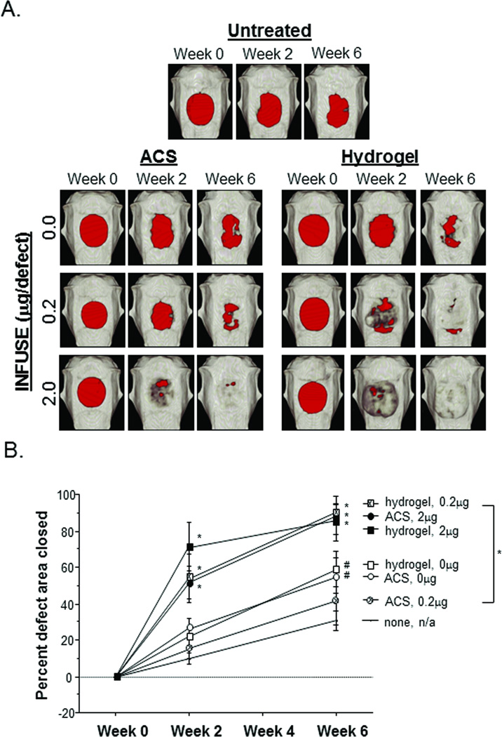
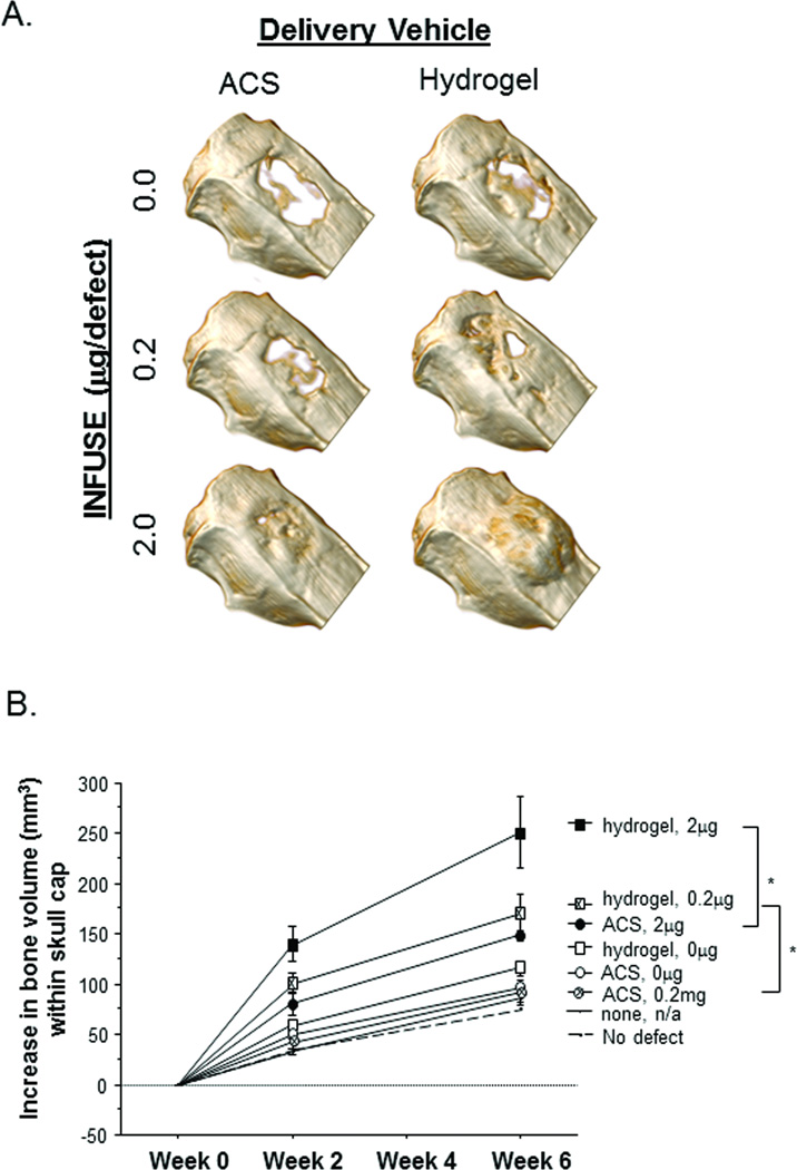
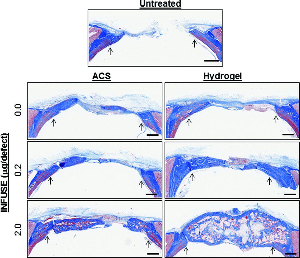
Medtronic's INFUSE Bone Graft provides surgeons with a potent tool for stimulating bone formation. Current delivery vehicles that rely on Absorbable Collagen Sponges (ACS) require excessive quantities of the active ingredient in INFUSE, recombinant human Bone Morphogenic Protein-2 (rhBMP2), to achieve physiologically relevant concentrations of the growth factor, driving up the cost of the product and increasing the likelihood of undesirable side effects in neighboring tissues. We demonstrate that a simple light-mediated, thiol-ene chemistry can be used to create an effective polymer delivery vehicle for rhBMP2, eliminating the use of xenographic materials and reducing the dose of rhBMP2 required to achieve therapeutic effects. Comprised entirely of synthetic components, this system entraps rhBMP2 within a biocompatible hydrogel scaffold that is degraded by naturally occurring remodeling enzymes, clearing the way for new tissue formation. When tested side-by-side with ACS in a critical-sized bone defect model in rats, this polymeric delivery system significantly increased bone formation over ACS controls.
Copyright © 2012 Orthopaedic Research Society.
Conflict of interest statement
The authors have no conflicts of interest to disclose regarding this work.
Figures
References
-
- Szpalski M, Gunzburg R. Recombinant human bone morphogenetic protein-2: a novel osteoinductive alternative to autogenous bone graft? Acta orthopaedica Belgica. 2005;71(2):133–148. - PubMed
-
- Valdes MA, Thakur NA, Namdari S, Ciombor DM, Palumbo M. Recombinant bone morphogenic protein-2 in orthopaedic surgery: a review. Archives of orthopaedic and trauma surgery. 2009;129(12):1651–1657. - PubMed
-
- Summary of Safety and Efficacy Data: Pre-Market Approval of InFUSE Bone Graft/LT-CAGE™ Lumbar Tapered Fusion Device. Food and Drug Administration; 2002.
-
- King GN. The importance of drug delivery to optimize the effects of bone morphogenetic proteins during periodontal regeneration. Current pharmaceutical biotechnology. 2001;2(2):131–142. - PubMed
Publication types
MeSH terms
Substances
Grants and funding
LinkOut - more resources
Full Text Sources
Other Literature Sources
