. 2012 Dec;32(24):5116-28.
doi: 10.1128/MCB.01034-12. Epub 2012 Oct 15.

Hippo signaling regulates pancreas development through inactivation of Yap

Affiliations

Hippo signaling regulates pancreas development through inactivation of Yap

Nicholas M George et al. Mol Cell Biol. 2012 Dec.

Abstract

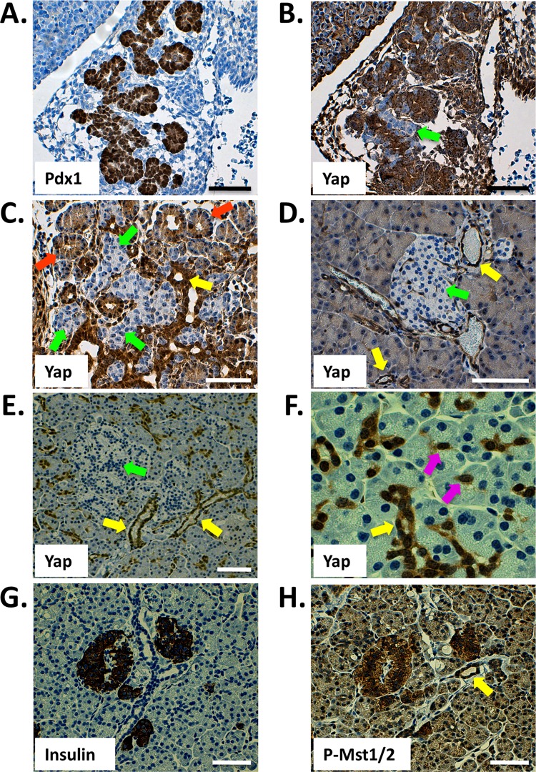
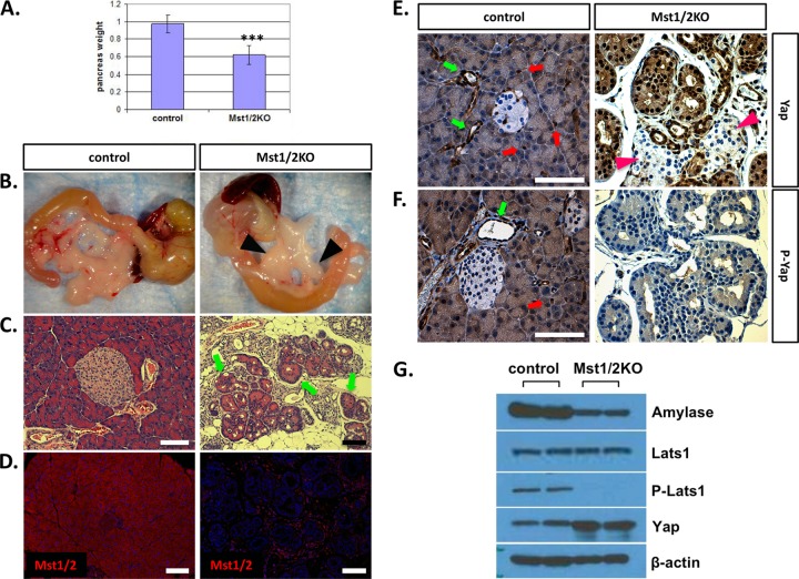
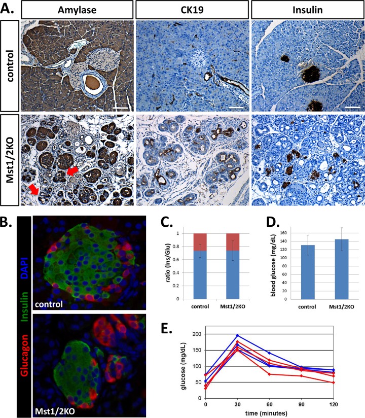
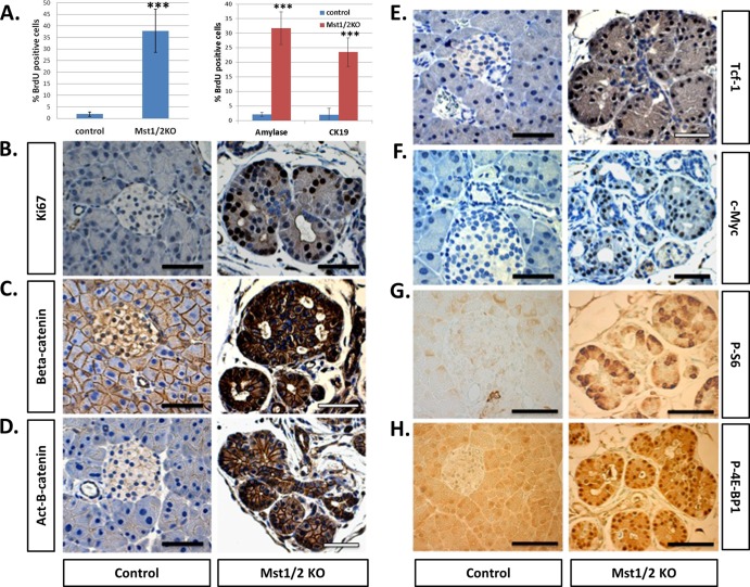
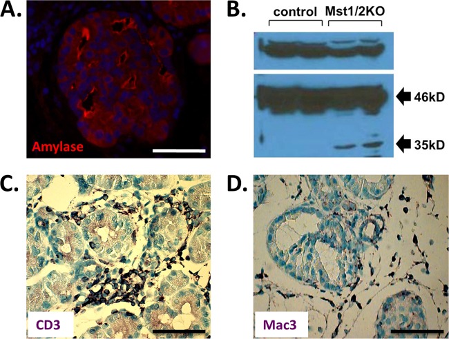
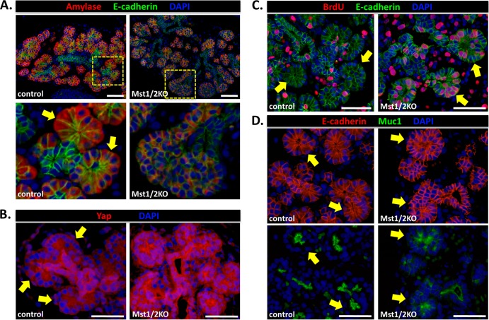
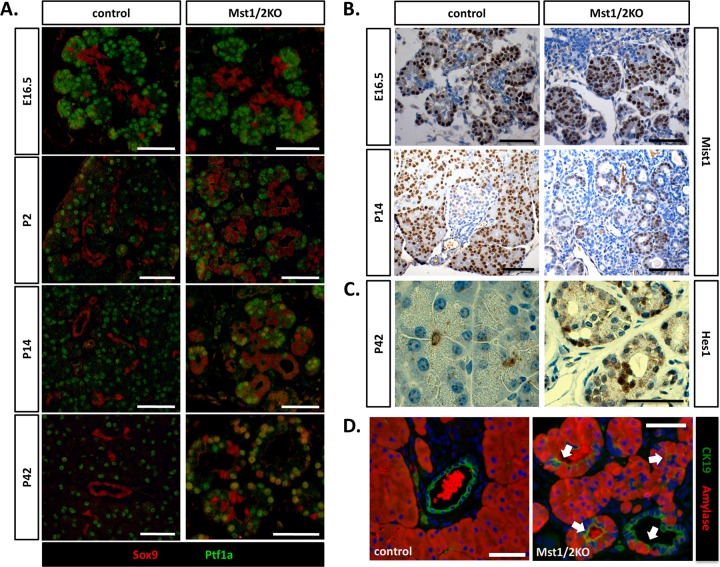
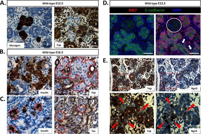
The mammalian pancreas is required for normal metabolism, with defects in this vital organ commonly observed in cancer and diabetes. Development must therefore be tightly controlled in order to produce a pancreas of correct size, cell type composition, and physiologic function. Through negative regulation of Yap-dependent proliferation, the Hippo kinase cascade is a critical regulator of organ growth. To investigate the role of Hippo signaling in pancreas biology, we deleted Hippo pathway components in the developing mouse pancreas. Unexpectedly, the pancreas from Hippo-deficient offspring was reduced in size, with defects evident throughout the organ. Increases in the dephosphorylated nuclear form of Yap are apparent throughout the exocrine compartment and correlate with increases in levels of cell proliferation. However, the mutant exocrine tissue displays extensive disorganization leading to pancreatitis-like autodigestion. Interestingly, our results suggest that Hippo signaling does not directly regulate the pancreas endocrine compartment as Yap expression is lost following endocrine specification through a Hippo-independent mechanism. Altogether, our results demonstrate that Hippo signaling plays a crucial role in pancreas development and provide novel routes to a better understanding of pathological conditions that affect this organ.


Figures

Fig 1
Fig 1
Immunohistochemical detection of Yap and active Mst1/2 in the mammalian pancreas. (A and B) Yap is broadly expressed throughout the Pdx1-positive E12.5 mouse pancreas (sequential sections). (C) Yap expression in the E16.5 pancreas. (D) Yap expression within the pancreas of the adult mouse (at 6 weeks) is largely confined to the ductal network, including terminal duct centroacinar cells, and is undetectable within islets. (E and F) Yap expression in the human pancreas (at 35 years) mirrors that of the mouse, with reactivity observable throughout the ductal network and absent from islets. (G and H) Active Hippo signaling, as determined by phosphorylation-specific Mst1/2 antibody, is present throughout the adult human pancreas. Highest levels of active Hippo signaling are found within islets (sequential sections). Arrows denote prospective or mature cell types, as follows: green, endocrine; orange, acinar; yellow, ducts; violet, centroacinar. Scale bar, 50 μm.
Fig 2
Fig 2
Conditional knockout of Mst1/2 in the mouse pancreas. Floxed Mst1/2 mice were crossed with the Pdx1-Cre strain to delete Mst1/2 throughout the developing pancreas. (A) Relative to controls, pancreas mass, as a percentage of total body weight, was significantly decreased in 6-week-old Mst1/2 KO offspring (***, P ≤ 0.01). (B) Loss of Mst1/2 leads to an atrophic phenotype characterized by immune cell infiltrate and prominent lymph nodes (arrowheads). (C) Transitional structures, consisting of both acinus-like and duct-like cells, are found throughout the 6-week Mst1/2 KO pancreas (arrows). (D) Efficient Cre-mediated recombination and excision of Mst1/2 as assessed by immunofluorescence detection of Mst1 and Mst2 using a dual-specificity antibody. (E and F) Both total Yap and the Ser127-phosphorylated form of Yap (P-Yap) are detected throughout the normal 6-week exocrine pancreas yet are absent from islet cells (red arrows, centroacinar cells; green arrows, ducts). Yap is nuclear localized and dephosphorylated in the 6-week Mst1/2 KO pancreas. Islands of endocrine cells (arrowheads) remain negative for Yap following Mst1/2 loss. (G) Canonical Hippo signaling regulates Yap phosphorylation and stability in the 6-week pancreas. Scale bar, 50 μm.
Fig 3
Fig 3
Pancreas-specific knockout of Mst1/2 leads to architectural defects in the exocrine and endocrine compartments yet has negligible effect on glucose homeostasis. (A) Mst1/2 knockout offspring lack the ordered arrangement of acinar cells observed in wild-type controls (both aged 6 weeks). Amylase positivity was detected in cells thought to be duct-like (arrows). Compared to controls, insulin-positive cells are highly dispersed in the Mst1/2 KOs. (B) Endocrine cell populations were characterized using immunofluorescent costaining of insulin and glucagon. Control mice contained normally appearing islets consisting of an insulin-positive interior surrounded by glucagon-positive exterior. Mst1/2 KO offspring harbored smaller islets consisting of cell clusters oftentimes expressing only a single hormone. (C) Ratios of insulin- to glucagon-expressing cells remained unchanged between control and knockout offspring (blue, insulin; red, glucagon). (D) Resting blood glucose levels reveal no significant difference between Mst1/2 KOs and controls (P = 0.35). (E) Fasting Mst1/2 KOs respond equally as well as controls following glucose challenge (red, Mst1/2 KO; blue, control). Scale bar, 50 μm.
Fig 4
Fig 4
Cell proliferation and signaling through the Wnt/β-catenin and mTOR pathways are robustly increased following pancreas-specific loss of Mst1/2 (6 weeks). (A) Total numbers of actively proliferating cells were quantified based on positivity for BrdU incorporation (left) as well as dual positivity of BrdU/amylase or BrdU/CK19 (right) (***, P ≤ 0.01). (B) Immunohistochemical detection of Ki67 in controls indicates very few proliferating cells, whereas Ki67-positive cells are readily observed in the Mst1/2 KOs. (C to H) The proproliferative Wnt/β-catenin and mTOR signaling pathways are activated following Mst1/2 loss in the pancreas. Scale bar, 50 μm.
Fig 5
Fig 5
Pancreas autodigestion counteracts cell proliferation. (A) Transitional structures display intense amylase staining along the cell surface of duct-like lumens suggestive of tissue autodigestion. (B) In contrast to controls, 6-week Mst1/2 KOs display intrapancreatic activation of carboxypeptidase A (CPA). Only the 46-kDa proform of CPA is evident in controls, whereas both the proform and the 35-kDa catalytically active form are present in the knockouts. (C and D) Mst1/2 KOs display a robust inflammatory infiltrate consisting of lymphocytes (e.g., CD3+ cells) and macrophage (Mac3+). Scale bar, 50 μm.
Fig 6
Fig 6
Hippo signaling becomes functionally active in the pancreas during the secondary transition. (A) Immunofluorescence analysis of control and Mst1/2 KO pancreas at E16.5. Loss of Hippo signaling had a trivial effect on the trunk region (E-cadherin) but a dramatic effect on the proacinar tip regions. Amylase staining is dramatically weaker in the Mst1/2 KOs, in line with greatly reduced cytoplasmic content. Tip cells in the knockouts were circular and lacked the “bottle-like” morphology displayed by cells in control mice (bottom panels, arrows). (B) Intense Yap immunoreactivity and nuclear localization within individual acini following Mst1/2 loss. Note the loss of Yap nuclear localization within individual acini of control pancreas. (C) In contrast to the acinar rosettes found in controls, acinar bundles in Mst1/2 KOs contain hyperproliferative cells. (D) Cell organization, as determined by staining with the luminal marker Muc1, is greatly disrupted following Mst1/2 loss. Scale bar, 50 μm.
Fig 7
Fig 7
Transitional structures are generated secondary to a normal exocrine differentiation program. (A) Differentiation within the Mst1/2 KO pancreas was monitored over time by analysis of Sox9 and Ptf1a expression. During embryonic pancreas development, Pft1a and Sox9 maintain their respective tip and trunk expression patterns regardless of Mst1/2 presence. Mixed differentiated, Sox9 and Ptf1a doubly positive cells were first observed at 2 weeks postpartum and increased to higher numbers by 6 weeks. (B) Robust expression of Mist1 in developing acini of both control and Mst1/2 KOs suggests normal exocrine differentiation. (C) Analysis of Hes1 expression suggests that transitional structures are not the result of an expanded centroacinar cell population. (D) In line with observation of frequent Sox9/Ptf1a doubly positive cells at 6 weeks, transitional structures contain cells of mixed exocrine status at this stage (arrows, amylase and CK19 copositive). Scale bar, 50 μm.
Fig 8
Fig 8
Yap expression is lost following endocrine specification in the pancreas. (A) Yap is undetectable within glucagon-positive endocrine clusters of the E12.5 mouse pancreas (sequential sections). (B and C) Both Yap and Taz are undetectable within insulin-expressing cells in the E16.5 mouse pancreas. (D) Direct correlation between endocrine specification and mitotic potential within the E12.5 mouse pancreas (the circle denotes a single endocrine cluster). Note cells negative for Ki67 along the length of the prospectively Ngn3-studded E-cadherin-positive trunk region (arrows). (E) Inverse correlation between Ngn3 and Yap expression within the E12.5 mouse pancreas (sequential sections; arrowheads represent individual Ngn3-positive/Yap-negative cells). Scale bar, 50 μm.

References

    1. Adler G, Hupp T, Kern HF. 1979. Course and spontaneous regression of acute pancreatitis in the rat. Virchows Arch. A Pathol. Anat. Histol. 382:31–47 - PubMed
    1. Bonner-Weir S, et al. 2004. The pancreatic ductal epithelium serves as a potential pool of progenitor cells. Pediatr. Diabetes 5:16–22 - PubMed
    1. Cai J, et al. 2010. The Hippo signaling pathway restricts the oncogenic potential of an intestinal regeneration program. Genes Dev. 24:2383–2388 - PMC - PubMed
    1. Camargo FD, et al. 2007. YAP1 increases organ size and expands undifferentiated progenitor cells. Curr. Biol. 17:2054–2060 - PubMed
    1. Criscimanna A, et al. 2011. Duct cells contribute to regeneration of endocrine and acinar cells following pancreatic damage in adult mice. Gastroenterology 141:1451–1462 - PMC - PubMed

Publication types

MeSH terms

LinkOut - more resources