. 2013 Jan;81(1):2-10.
doi: 10.1128/IAI.00666-12. Epub 2012 Oct 15.

Vitamin D rescues impaired Mycobacterium tuberculosis-mediated tumor necrosis factor release in macrophages of HIV-seropositive individuals through an enhanced Toll-like receptor signaling pathway in vitro

Affiliations

Vitamin D rescues impaired Mycobacterium tuberculosis-mediated tumor necrosis factor release in macrophages of HIV-seropositive individuals through an enhanced Toll-like receptor signaling pathway in vitro

Asha Anandaiah et al. Infect Immun. 2013 Jan.

Abstract

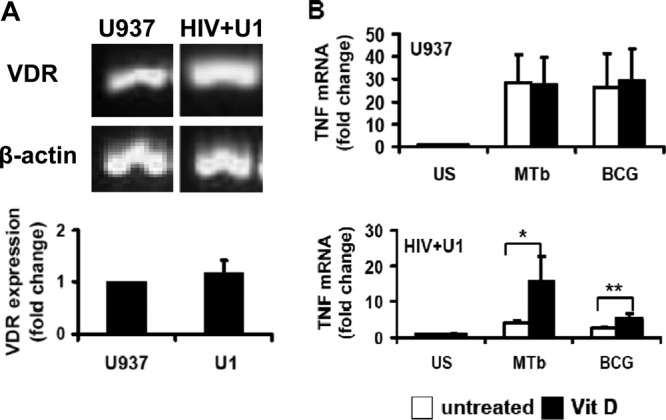
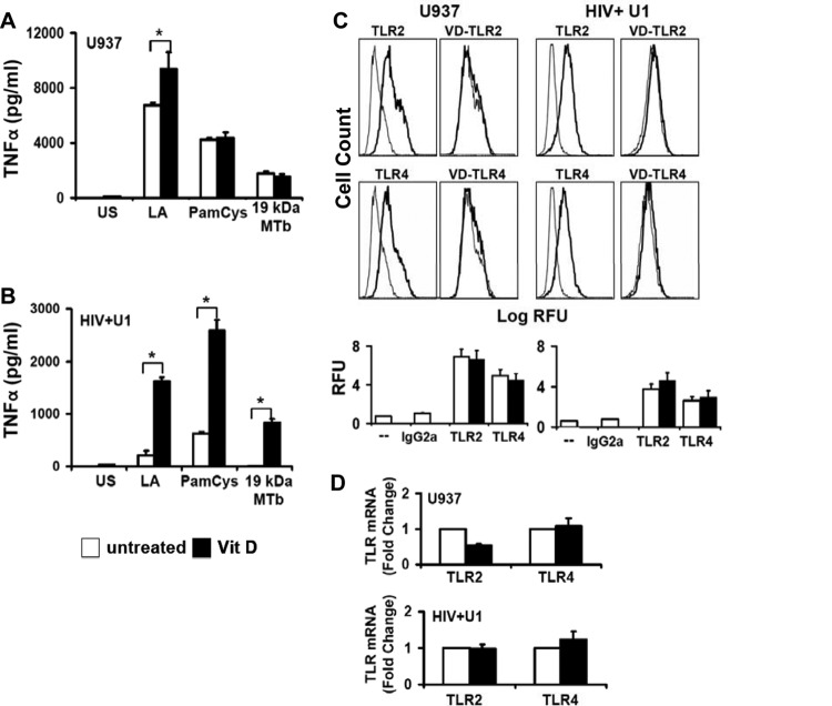
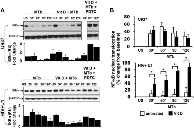
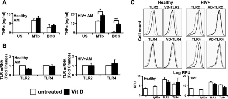
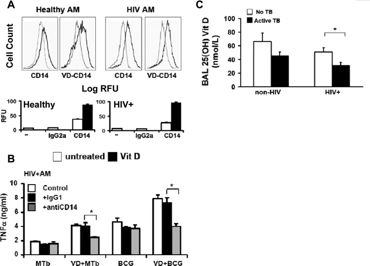
Mycobacterium tuberculosis disease represents an enormous global health problem, with exceptionally high morbidity and mortality in HIV-seropositive (HIV(+)) persons. Alveolar macrophages from HIV(+) persons demonstrate specific and targeted impairment of critical host cell responses, including impaired M. tuberculosis-mediated tumor necrosis factor (TNF) release and macrophage apoptosis. Vitamin D may promote anti-M. tuberculosis responses through upregulation of macrophage NO, NADPH oxidase, cathelicidin, and autophagy mechanisms, but whether vitamin D promotes anti-M. tuberculosis mechanisms in HIV(+) macrophages is not known. In the current study, human macrophages exposed to M. tuberculosis demonstrated robust release of TNF, IκB degradation, and NF-κB nuclear translocation, and these responses were independent of vitamin D pretreatment. In marked contrast, HIV(+) U1 human macrophages exposed to M. tuberculosis demonstrated very low TNF release and no significant IκB degradation or NF-κB nuclear translocation, whereas vitamin D pretreatment restored these critical responses. The vitamin D-mediated restored responses were dependent in part on macrophage CD14 expression. Importantly, similar response patterns were observed with clinically relevant human alveolar macrophages from healthy individuals and asymptomatic HIV(+) persons at high clinical risk of M. tuberculosis infection. Taken together with the observation that local bronchoalveolar lavage fluid (BALF) levels of vitamin D are severely deficient in HIV(+) persons, the data from this study demonstrate that exogenous vitamin D can selectively rescue impaired critical innate immune responses in vitro in alveolar macrophages from HIV(+) persons at risk for M. tuberculosis disease, supporting a potential role for exogenous vitamin D as a therapeutic adjuvant in M. tuberculosis infection in HIV(+) persons.


Figures

Fig 1
Fig 1
1,25D3 rescues M. tuberculosis-mediated TNF release in HIV+ human macrophages. Differentiated U937 (A) and HIV+ U1 (B and C) macrophages were incubated with irradiated virulent M. tuberculosis (MTb) (MOI of 10:1 for 24 h) in the presence or absence of 1,25D3 (VD) pretreatment (24 h). TNF in cell culture supernatants was measured by ELISA (R&D). Figures are representative of individual experiments with similar results (n = 6). Quantitative data represent means ± standard errors of the means (SEM). US, unstimulated. *, P < 0.05.
Fig 2
Fig 2
1,25D3 enhances TNF transcription in HIV+ human macrophages. (A) RT-PCR and real-time PCR for VDR were performed on total RNAs from differentiated U937 and HIV+ U1 macrophages (n = 4). (B) Differentiated U937 and HIV+ U1 cells were incubated with M. tuberculosis or BCG (M. bovis) (MOI, 10:1) for 3 h in the presence or absence of 1,25D3 pretreatment (24 h). TNF mRNA was measured by real-time PCR (n = 4). Quantitative data represent means ± SEM. *, P < 0.05; **, P < 0.01.
Fig 3
Fig 3
1,25D3 increases TLR signaling but not TLR expression in HIV+ U1 macrophages. (A and B) Differentiated human U937 and HIV+ U1 macrophages were incubated with the TLR ligands lipid A (LA) (for TLR4), PamCys (for TLR2/1), and 19-kDa lipoprotein from M. tuberculosis (19 kDa MTb; 1 μg/ml) (for TLR2/1) for 24 h in the presence or absence of 1,25D3 pretreatment. Cell-free culture supernatants were assayed for TNF by ELISA (n = 3). (C) Differentiated U937 and HIV+ U1 macrophages were incubated for 24 h in the presence or absence of 1,25D3 pretreatment and then stained with PE-labeled anti-TLR antibodies or isotype control antibody. Surface expression was measured by flow cytometry. Left panels show isotype control (gray lines)- and TLR (black lines)-labeled cells; right panels show TLR-labeled cells (gray lines) and 1,25D3-treated TLR-labeled cells (black lines). Representative histograms for independent experiments with similar results (n = 3) are shown. (D) Specific TLR2 and TLR4 mRNAs were detected by real-time PCR (n = 3). Quantitative data represent means ± SEM. *, P < 0.05.
Fig 4
Fig 4
1,25D3 upregulates NF-κB signaling in HIV+ human macrophages. (A) Differentiated U937 and HIV+ U1 macrophages were incubated with M. tuberculosis (MOI, 10:1) for 0 to 120 min in the presence or absence of 1,25D3 and PDTC. Cell lysates were resolved by Western blotting using a specific antibody to IκBα. Representative blots for three independent experiments with similar results are shown. Quantitative densitometric analyses of IκBα bands are displayed directly beneath the blots. (B) NF-κB nuclear translocation in nuclear extracts was measured by ELISA (n = 3). Quantitative data represent means ± SEM. *, P < 0.05.
Fig 5
Fig 5
1,25D3 induces CD14 expression in human macrophages. (A) Differentiated U937 and HIV+ U1 macrophages were incubated for 24 h in the presence or absence of 1,25D3 pretreatment and then stained with PE-labeled anti-CD14 antibody or isotype control antibody. Surface expression was measured by flow cytometry. Left panels show isotype control (gray lines)- and CD14 (black lines)-labeled cells; right panels show CD14-labeled cells (gray lines) and 1,25D3-treated CD14-labeled cells (black lines). Representative histograms from individual experiments with similar results (n = 3) are shown. (B and C) Differentiated U937 (B) and HIV+ U1 (C) macrophages were treated with M. tuberculosis (MOI, 10:1) or LPS (1 μg/ml) in the presence or absence of vitamin D pretreatment (24 h) and the indicated antibodies. TNF in cell culture supernatants was measured by ELISA (R&D). The data are representative of three individual experiments with similar results. Quantitative data represent means ± SEM. *, P < 0.05.
Fig 6
Fig 6
1,25D3 rescues M. tuberculosis-mediated TNF release in human alveolar macrophages from HIV+ persons. (A) AMs from healthy (n = 6) and HIV+ (n = 2) persons were incubated with M. tuberculosis or BCG (MOI of 10:1 for 24 h) in the presence or absence of 1,25D3 pretreatment (24 h). TNF in cell culture supernatants was measured by ELISA (R&D). (B) Specific TLR2 and TLR4 mRNAs were detected by real-time PCR (n = 3). (C) AMs were incubated for 24 h in the presence or absence of 1,25D3 pretreatment and then stained with PE-labeled anti-TLR or isotype control antibody. Surface expression was measured by flow cytometry. Left panels show isotype control (gray lines)- and receptor (black lines)-labeled cells; right panels show receptor-labeled cells (gray lines) and 1,25D3-treated receptor-labeled cells (black lines). Representative histograms for individual experiments with similar results (n = 3 for healthy individuals and 2 for HIV+ individuals) are shown. Quantitative data represent means ± SEM. *, P < 0.05; **, P < 0.01.
Fig 7
Fig 7
1,25D3 rescue of M. tuberculosis-mediated TNF release in human alveolar macrophages from HIV+ persons is dependent on CD14. (A) AMs were incubated for 24 h in the presence or absence of 1,25D3 pretreatment and then stained with PE-labeled anti-CD14 or isotype control antibody. Surface expression was measured by flow cytometry. Left panels show isotype control (gray lines)- and receptor (black lines)-labeled cells; right panels show receptor-labeled cells (gray lines) and 1,25D3-treated receptor-labeled cells (black lines) (n = 3). (B) HIV+ AMs were treated with M. tuberculosis (MOI of 0.25:1), BCG (MOI of 10:1), or LPS (1 μg/ml) in the presence or absence of 1,25D3 pretreatment (24 h) and the indicated antibodies. TNF in cell culture supernatants was measured by ELISA (n = 2). (C) 25D3 levels were measured in the cell-free BALF of healthy and HIV-infected Indian patients with and without active M. tuberculosis infection (for HIV M. tuberculosis individuals, n = 38; for HIV M. tuberculosis+ individuals, n = 35; for HIV+ M. tuberculosis individuals, n = 12; and for HIV+ M. tuberculosis+ individuals, n = 17) by ELISA. Quantitative data represent means ± SEM. *, P < 0.05.

References

    1. Anandaiah A, Dheda K, Keane J, Koziel H, Moore DA, Patel NR. 2011. Novel developments in the epidemic of human immunodeficiency virus and tuberculosis coinfection. Am. J. Respir. Crit. Care Med. 183:987–997 doi:10.1164/rccm.201008-1246CI - DOI - PMC - PubMed
    1. WHO 2010. Global tuberculosis control: WHO report 2010. WHO, Geneva, Switzerland
    1. Moreno S, Baraia-Etxaburu J, Bouza E, Parras F, Perez-Tascon M, Miralles P, Vicente T, Alberdi JC, Cosin J, Lopez-Gay D. 1993. Risk for developing tuberculosis among anergic patients infected with HIV. Ann. Intern. Med. 119:194–198 - PubMed
    1. Sonnenberg P, Glynn JR, Fielding K, Murray J, Godfrey-Faussett P, Shearer S. 2005. How soon after infection with HIV does the risk of tuberculosis start to increase? A retrospective cohort study in South African gold miners. J. Infect. Dis. 191:150–158 - PubMed
    1. Williams BG, Dye C. 2003. Antiretroviral drugs for tuberculosis control in the era of HIV/AIDS. Science 301:1535–1537 - PubMed

Publication types

MeSH terms