. 2012 Oct 22;209(11):2065-77.
doi: 10.1084/jem.20112272. Epub 2012 Oct 15.

Deletion and anergy of polyclonal B cells specific for ubiquitous membrane-bound self-antigen

Affiliations

Deletion and anergy of polyclonal B cells specific for ubiquitous membrane-bound self-antigen

Justin J Taylor et al. J Exp Med. .

Abstract

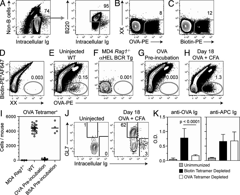
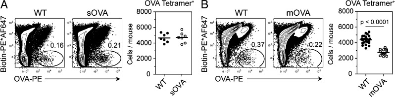
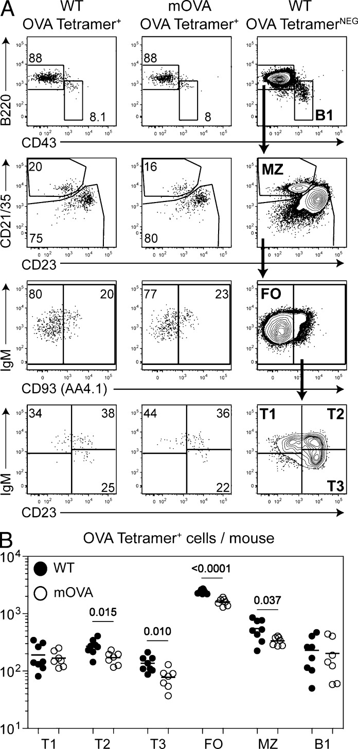
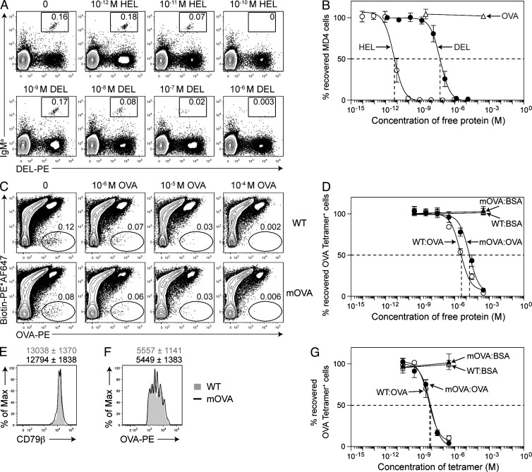
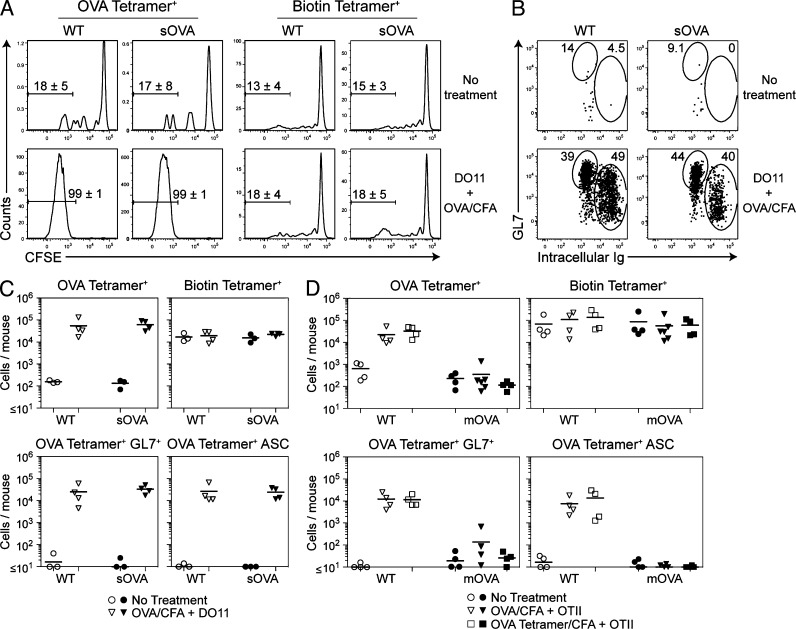
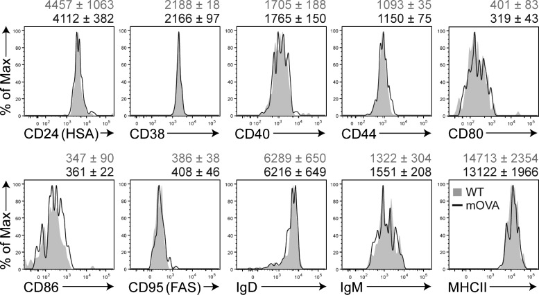
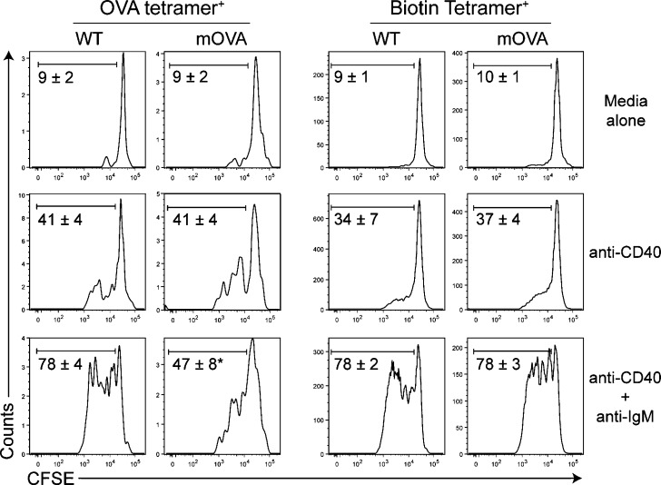
B cell tolerance to self-antigen is critical to preventing antibody-mediated autoimmunity. Previous work using B cell antigen receptor transgenic animals suggested that self-antigen-specific B cells are either deleted from the repertoire, enter a state of diminished function termed anergy, or are ignorant to the presence of self-antigen. These mechanisms have not been assessed in a normal polyclonal repertoire because of an inability to detect rare antigen-specific B cells. Using a novel detection and enrichment strategy to assess polyclonal self-antigen-specific B cells, we find no evidence of deletion or anergy of cells specific for antigen not bound to membrane, and tolerance to these types of antigens appears to be largely maintained by the absence of T cell help. In contrast, a combination of deleting cells expressing receptors with high affinity for antigen with anergy of the undeleted lower affinity cells maintains tolerance to ubiquitous membrane-bound self-antigens.


Figures

Figure 1.
Figure 1.
Analysis of OVA-specific B cells with an OVA tetramer. (A) B cells were identified by flow cytometry in pooled spleen and LN samples as cells that bound an antibody specific for Ig (binds both intracellular and surface Ig), but not a cocktail of antibodies specific for Thy1.2 (or CD4 and CD8), CD11c, Gr-1, or F4/80. Within the population, cells that neither expressed B220 or high levels of intracellular Ig were excluded. Representative flow cytometric analysis of gated B cells in a fraction enriched using anti-PE magnetic microbeads after staining with OVA-PE tetramer (B), Biotin-PE tetramer (C), Biotin-PE*AF647 tetramer (D), or both OVA-PE tetramer and Biotin-PE*AF647 tetramer (E–H). XX indicates an empty PE*AF647 (B and C) or PE (D) channel. In H, mice were injected s.c. with 1.4 nmol OVA emulsified in CFA 18 d before analysis. In G and I, cells were incubated with 300 µM of monomeric OVA or BSA beginning 20 min before tetramer labeling. (I) Combined data from 20 experiments showing the number of OVA tetramer+ B cells in individual mice. The bar indicates mean. (J) Representative flow cytometric analysis of GL7 and intracellular Ig expression on gated OVA-specific B cells found in naive and mice injected s.c. with 1.4 nmol OVA emulsified in CFA. The flow cytometry plots shown in A–G and J are representative of at least three independent experiments. (K) Combined data from 2 experiments showing serum anti-OVA or anti-APC IgG+IgM measured 10 d after s.c. injection of Rag1−/− with 5.6 nmol of OVA and 0.1 nmol APC in CFA. Before injection, Rag1−/− received the entire fraction of spleen and LN cells that flowed through the column of a Biotin-PE*AF647 tetramer enrichment alone (Biotin tetramer depleted) or OVA-PE tetramer + Biotin-PE*AF647 tetramer enrichments (OVA tetramer depletion). Bars represent the mean ± SD (n = 5–9), and the p-value was established using an unpaired two-tailed Student’s t test.
Figure 2.
Figure 2.
Enumeration of OVA-specific B cells in OVA-expressing animals. Representative flow cytometric analysis and total cell number of gated B cells in a fraction enriched using anti-PE magnetic microbeads after staining with both OVA-PE tetramer and Biotin-PE*AF647 tetramer from mice expressing secreted OVA (sOVA; A) or membrane-bound OVA (mOVA; B) mice. Corresponding WT mice are shown as a control. Data are combined from 7–15 experiments and represent the number of cells found in an individual mouse. The line indicates the mean (n = 7–26), and the p-value was established using an unpaired two-tailed Student’s t test.
Figure 3.
Figure 3.
B cell subpopulation analysis of OVA-specific B cells from WT and mOVA-expressing animals. (A) Representative flow cytometric gating strategy for subpopulation analysis of OVA tetramer+ and OVA TetramerNEG B cells (CD19+ Thy1.2 CD11c Gr-1 F4/80) in WT and mOVA mice. FO, mature FO; MZ, marginal zone. (B) Combined data from three experiments showing the total number of OVA tetramer+ B cells within each subpopulation from individual mice (n = 8). The line indicates the mean, and the values above data points are p-values established using an unpaired two-tailed Student’s t test.
Figure 4.
Figure 4.
Measuring the median affinity of polyclonal OVA-specific B cells. Spleen and LNs from four mice were pooled and split equally into eight tubes and incubated with the noted concentration of monomeric antigen before tetramer enrichment and flow cytometric analysis. In A and B, ∼16,000 MD4 Rag1−/− B cells were added to a WT B6 sample before antigen incubation (HEL, DEL, or OVA) and enriched using a DEL tetramer. (A) Representative flow cytometric analysis showing the detection of MD4 B cells as IgMa+ DEL tetramer+ cells. (B) Combined data from 6 experiments showing the percentage of MD4 cells recovered compared with the number of cells detected in a sample in which no monomeric antigen competitor was added. Each data point represents the mean ± SEM (n = 3–7). (C) Representative flow cytometric analysis showing the detection of OVA tetramer+ B cells from WT and mOVA mice with or without antigen (OVA or BSA) preincubation. (D) Combined data from three experiments showing the percentage of OVA tetramer+ B cells recovered compared with the number of cells detected in a sample in which no monomeric antigen competitor was added. Each data point represents the mean ± SEM (n = 3–7). (E) Representative flow cytometric analysis of surface Igβ (CD79b) expression by OVA Tetramer+ B cells from mOVA and WT mice. The numbers above the plots represent the mean fluorescence intensity ± SD (n = 5–6) of cells from individual WT (gray) and mOVA (black) animals from 2 experiments. (F) Representative flow cytometric analysis of the level of OVA tetramer+ on OVA tetramer+ B cells from mOVA and WT mice. Numbers above represent the mean fluorescence intensity ± SD (n = 11–12) of OVA tetramer from individual mice from 4 separate experiments. (G) Same as D, except samples were incubated with APC-conjugated OVA or BSA tetramers before labeling and enrichment with Biotin-PE*AF647 and OVA-PE tetramers. Each data point represents the mean ± SEM (n = 3).
Figure 5.
Figure 5.
Functional capabilities of OVA-specific B cells found in OVA-expressing animals. MACS-purified B cells from sOVA, mOVA, or WT control mice were CFSE-labeled and adoptively transferred into Rag1−/− recipients. Some mice also received 10,000 syngeneic OVA-specific CD4+ helper T cells (DO11 Rag2−/− or OTII Rag1−/−) and s.c. injection of 5.5 nmol of OVA or 1.4 nmol OVA tetramer in CFA 7 d before analysis. After OVA-PE tetramer and Biotin-PE*AF647 tetramer labeling and enrichment, gated OVA tetramer+ and Biotin tetramer+ cells were analyzed for CFSE dilution (A) or GL7 and intracellular Ig expression (B). Representative plots are shown and the numbers represent the mean ± SD (n = 3–4) percentage of CFSELOW cells found in individual mice from 2 combined experiments. Combined data from three to four experiments showing the total number of OVA tetramer+, Biotin tetramer+, OVA tetramer+ GL7+, and OVA tetramer+ ASC (IgHI) B cells in individual recipients of sOVA (C) or mOVA (D) B cells. Corresponding WT mice are shown as a control. The line indicates the mean (n = 3–6).
Figure 6.
Figure 6.
Surface marker expression by OVA-specific B cells found in mOVA-expressing animals. Representative flow cytometric analysis of the expression of MHC class II (MHCII), CD95 (FAS), CD38, CD40, CD44, CD80, CD86, CD24 (HSA), IgM, and IgD by mature FO OVA tetramer+ B cells from mOVA and WT mice gated as shown in Fig. 3. The numbers above the plots represent the mean fluorescence intensity ± SD (n = 3–8) from individual WT (gray) and mOVA (black) animals combined from 2–4 experiments per marker.
Figure 7.
Figure 7.
In vitro response of OVA-specific B cells from WT and mOVA-expressing animals. MACS-purified B cells from mOVA or WT control mice were cultured in vitro in the presence of 1 µg/ml αCD40, 1 µg/ml αCD40, and 5 µg/ml αIgM, or media alone for 72 h before tetramer labeling and enrichment and analysis of CFSE dilution. The numbers on the plot represent the mean percentage (±SD) of CFSELOW cells found in three separate experiments. *, P = 0.038 compared with the percentage of CFSELOW cells found in the OVA tetramer+ population from WT mice stimulated with αCD40 and αIgM.
Figure 8.
Figure 8.
Analysis of GPI-specific B cells in an adoptive transfer model of arthritis. (A) Representative flow cytometric analysis from 15 experiments of gated B cells in the GPI-PE tetramer and C5a-PE*AF647 tetramer-enriched fraction of Tcra−/− B6.I-Ab/g7 mice with and without adoptive transfer of GPI-specific CD4+ helper T cells (KRN) 7 d before analysis. (B) Combined data from 15 experiments showing the number of GPI tetramer+ B cells in individual HEL-specific MD4 Rag1−/−, B6.I-Ab/g7, Tcra−/− B6.I-Ab/g7, and Tcra−/− B6.I-Ab/g7 mice 7 d after transfer of KRN cells. The line indicates the mean (n = 3–26). (C) Combined data from three experiments showing the number of GPI tetramer+ B cells of each subpopulation identified as shown in Fig. 3. (D) Combined data from four experiments showing the percentage of GPI Tetramer+ B cells recovered in the presence of monomeric GPI or OVA competitor as compared with the number of cells detected in a sample in which no competitor antigen was added. Each data point represents the mean ± SEM (n = 5). (E, left) Pooled data from 2 experiments showing the arthritis clinical disease activity scores (max = 12) of individual Tcra−/− B6.I-Ab/g7 mice (n = 4) and (right) mean (n = 5; ± SEM) serum anti-GPI IgG1 measured on the days indicated after adoptive transfer of KRN CD4+ helper T cells. Serum anti-GPI IgG1 antibody levels reflect the optical density measured at 1:900 dilution of serum. (F) Representative flow cytometric analysis from 5 experiments showing GL7 and intracellular Ig expression on gated GPI tetramer+ B cells 7 d after transfer of KRN cells. (G) Representative flow cytometric analysis from 3 experiments showing surface IgG1 expression on GL7+ and intracellular IgHI GPI tetramer+ populations 7 d after transfer of KRN cells. (H) Pooled data from 3 experiments showing the arthritis clinical disease activity scores (max = 12) 12 d after the transfer of KRN CD4+ helper T cells into NOD Rag1−/− IL2rγ−/− recipient mice that received FACS-purified GPI tetramer+ or C5a tetramer+ B cells. Each data point indicates an individual mouse (n = 5–7), and the bar indicates the mean. The p-values were established using an unpaired two-tailed Student’s t test. (I) Arthritis clinical disease activity scores from the recipients of GPI tetramer+ B cells from H plotted against the total number of GPI tetramer+ B cells in the recipient animals 12 d after the transfer of KRN CD4+ helper T cells.

References

    1. Abbas A.K., Lohr J., Knoechel B. 2007. Balancing autoaggressive and protective T cell responses. J. Autoimmun. 28:59–61 10.1016/j.jaut.2007.02.002 - DOI - PMC - PubMed
    1. Adelstein S., Pritchard-Briscoe H., Anderson T.A., Crosbie J., Gammon G., Loblay R.H., Basten A., Goodnow C.C. 1991. Induction of self-tolerance in T cells but not B cells of transgenic mice expressing little self-antigen. Science. 251:1223–1225 10.1126/science.1900950 - DOI - PubMed
    1. Barnden M.J., Allison J., Heath W.R., Carbone F.R. 1998. Defective TCR expression in transgenic mice constructed using cDNA-based alpha- and beta-chain genes under the control of heterologous regulatory elements. Immunol. Cell Biol. 76:34–40 10.1046/j.1440-1711.1998.00709.x - DOI - PubMed
    1. Borrero M., Clarke S.H. 2002. Low-affinity anti-Smith antigen B cells are regulated by anergy as opposed to developmental arrest or differentiation to B-1. J. Immunol. 168:13–21 - PubMed
    1. Cambier J.C., Gauld S.B., Merrell K.T., Vilen B.J. 2007. B-cell anergy: from transgenic models to naturally occurring anergic B cells? Nat. Rev. Immunol. 7:633–643 10.1038/nri2133 - DOI - PMC - PubMed

Publication types

MeSH terms