. 2012 Dec 6;120(24):4802-11.
doi: 10.1182/blood-2012-08-449744. Epub 2012 Oct 15.

IGHV-unmutated and IGHV-mutated chronic lymphocytic leukemia cells produce activation-induced deaminase protein with a full range of biologic functions

Affiliations

IGHV-unmutated and IGHV-mutated chronic lymphocytic leukemia cells produce activation-induced deaminase protein with a full range of biologic functions

Piers E M Patten et al. Blood. .

Abstract

Clonal evolution occurs during the course of chronic lymphocytic leukemia (CLL) and activation-induced deaminase (AID) could influence this process. However, this possibility has been questioned in CLL because the number of circulating AID mRNA(+) cells is exceedingly low; synthesis of AID protein by blood CLL cells has not been demonstrated; the full range of AID functions is lacking in unmutated CLL (U-CLL), and no prospective analysis linking AID expression and disease severity has been reported. The results of the present study show that circulating CLL cells and those within secondary lymphoid tissues can make AID mRNA and protein. This production is related to cell division because more AID mRNA was detected in recently divided cells and AID protein was limited to the dividing fraction and was up-regulated on induction of cell division. AID protein was functional because AID(+) dividing cells exhibited more double-stranded DNA breaks, IGH class switching, and new IGHV-D-J mutations. Each of these actions was documented in U-CLL and mutated CLL (M-CLL). Furthermore, AID protein was associated with worse patient outcome and adverse cytogenetics. We conclude that the production of fully functional AID protein by U-CLL and M-CLL cells could be involved in clonal evolution of the disease.


Figures
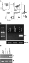
Figure 1
Figure 1
AID mRNA expression relates to recent cell division. (A) CD5+CD19+ cells were sorted into recently divided (CD5+CD19+CD23BrCD11a+CXCRDim), intermediate (CD5+CD19+CD23ModCD11a+/−CXCR4Mod), and resting (CD5+CD19+ CD23DimCD11aCXCR4Br) fractions before analysis of mRNA transcripts for AID. (B) Representative agarose gel of mRNA for AID and β-actin in sorted fractions. Both full-length and a spliced AID transcripts can be seen. (C) In this example, CLL patient-specific μ transcripts were found in all 3 cell fractions by RT-PCR, but switched γ transcripts were only detected in the proliferative fraction. DNA sequencing confirmed that the γ and μ transcripts were of patient origin.
Figure 2
Figure 2
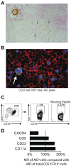
CLL cells in infiltrated LNs express AID protein. (A) Representative low-power (100× original magnification) and high-power (600× original magnification, inset) views of AID+ cells in an infiltrated CLL LN. Scattered AID+ cells (black arrows), which are larger than the majority of cells, are present and have the morphology of paraimmunoblasts (inset). (B) Representative high-power confocal photomicrograph of CLL LN (600× original magnification) showing cells expressing the CLL marker CD23 (red). Several cells are Ki-67+ (blue). An AID+Ki-67+ cell (green) is indicated by the white arrow. (C) Flow cytometry of dispersed LN cells. The percentage of AID+ cells was determined by comparison of samples with and without the use of a peptide that specifically blocks binding of the AID Ab. At least 1% CD5+CD19+ were AID+. (D) Graph showing the mean fluorescence intensity (MFI) of CXCR4, CD5, CD23, and CD11a in AID+ cells compared with the total CD5+CD19+ sorted population using the same gating as in panel C.
Figure 3
Figure 3
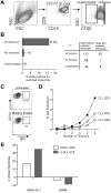
In vitro–activated CLL PBMCs express AID protein. (A) Percentage of AID+ cells within the total CD5+CD19+ population derived from 16 cocultures of CLL PBMCs and CD32-transfected fibroblasts stimulated with CD40 + IL-4 for 72 hours. (B) Comparison of the presence or absence of AID mRNA prestimulation with the percentage CD5+CD19+ cells expressing AID protein 72 hours poststimulation. Means ± SEM of 16 samples are shown. *P = .01 by unpaired t test. (C) Detection of AID protein (white fill) compared with rat IgG2b isotype control mAb (gray fill) by FACS on CD5+CD19+ cells stimulated in the CD40 + IL-4 system at 0, 72, and 168 hours. (D) Confocal photomicrograph of CD23+ cells (red) from the CD40 + IL-4 system visualized at 168 hours demonstrating AID protein (green) localized only in the cytoplasm (replicating protein A-blue (RPA) is used as a nuclear stain). Original magnification was 600×. (E) Representative FACS plots of AID staining on CD5+CD19+ cells derived from unstimulated and CD40 + IL-4–stimulated, CFSE-labeled CLL PBMCs after 7 days. Percentages of AID+ cells in the total CD5+CD19+ population are shown. (F) Three patterns of AID up-regulation were observed after CD5+CD19+ cells were cultured for 7 days in the CD40 + IL-4 system: (I) no up-regulation (M-CLL922, M-CLL1227, and M-CLL1232), (II) up-regulation with each division cycle (M-CLL1082, M-CLL1201, U-CLL1238, M-CLL1252, and M-CLL1299), and (III) up-regulation after ≤ 2 cycles (M-CLL797, U-CLL976, and U-CLL1278). (G) Graphs comparing the percentage AID+CD5+CD19+ cells from 5 cultures at 7 and 14 days. Colors denote individual patient samples: purple: U-CLL1278, blue: M-CLL1082, green: M-CLL1299, black: M-CLL1252, and red: M-CLL922).
Figure 4
Figure 4
CLL cells that divided and up-regulated AID protein exhibited more dsDNA breaks. (A) Confocal photomicrographs comparing CLL PBMCs stimulated in the CD40 + IL-4 system with unstimulated cells cocultured only with CD32-transfected fibroblasts. Original magnification was 630×. (B) Quantitative colocalization of CFSE intensity (x axis) and anti-pH2A.X staining (y-axis) on CD23+ cells derived from stimulated cultures. The shaded area (gray) represents the range of pH2A.X intensity derived from unstimulated cells, all of which had a CFSE intensity of at least 256 pixels; numbers denote the quantity of cells present in each of the 4 quadrants. ***P < .0001 by Fisher exact test. (C) Graph showing the change in AID mean fluorescence intensity (MFI) identified in CD5+CD19+ cells of the same 3 samples in panel B as determined by flow cytometry.
Figure 5
Figure 5
CLL cells that divided and up-regulated AID protein exhibited CSR and de novo SHM. (A) Sort strategy to obtain 20-cell and 1-cell aliquots of CD5+CD19+ cells from CLL PBMCs after up to 14 days of culture. (B) Comparison of the percentages and numbers of 20-cell/well aliquots that yielded α and γ (switched) CLL patient–specific IGHV-D-J transcripts from divided, undivided, and unstimulated CD5+CD19+ populations after 14 days of culture. ***P = .0002 by χ2 test. (C) Representative FACS plots showing minimal surface IgG expression by CD5+CD19+ cells that had not divided after 14 days of stimulation compared with at least a 10-fold higher expression by multiply divided cells. (D) Comparison of the division number with the percentage of surface IgG expression by CD5+CD19+ cells derived from stimulated CLL PBMCs after 14 days of culture. (E) Change in number of unique mutated subclones identified by NGS from cells stimulated in the CD40 + IL-4 system compared with prestimulated samples. The change in unique subclone count is subdivided into mutations in IGHVDJ and IGHM; a negative number was generated for IGHM in both cases because less unique subclones were found in this region after stimulation compared with prestimulated cells.
Figure 6
Figure 6
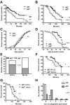
AID+ CLL is correlated with an increased number of cytogenetic aberrations and worse clinical outcomes. Clinical data and prognostic factors for AID+ and AID CLL patients were compared using the Kaplan-Meier method. (A) TFT curves comparing AID+ and AID CLL patients (n = 58 and n = 55, respectively) were significantly different. **P = .0023. (B) OS curve of AID+ CLL patients (n = 57) differed significantly from that of AID patients (n = 55) ***P < .0001. (C) Age of CLL diagnosis curves of AID+ and AID CLL patients were not significantly different (n/s). (D) Age of death curve for AID+ CLL patients differed significantly from that of AID patients. ***P < .0001. (E) U-CLL patients were more frequently AID+ (38/55), whereas M-CLL patients were more frequently AID (54 of 66). ***P = .0001. (F) OS curve of AID+ M-CLL patients differed significantly from that of AID M-CLL patients. *P = .0264. (G) OS curves of AID+ and AID U-CLL patients were not significantly different (n/s). (H) AID+ patients had increased numbers of aberrations at 7 commonly tested cytogenetic loci (17p13.1, 11q22.3, 12-CEN, 13q34, 11q13, 14q32, and 6q23.3) compared with AID patients (median = 1 vs 0). Distribution of the numbers of aberrations was significantly different between the AID+ and AID groups. ***P = .0009.

References

    1. Chiorazzi N, Rai KR, Ferrarini M. Chronic lymphocytic leukemia. N Engl J Med. 2005;352(8):804–815. - PubMed
    1. Shanafelt TD, Witzig TE, Fink SR, et al. Prospective evaluation of clonal evolution during long-term follow-up of patients with untreated early-stage chronic lymphocytic leukemia. J Clin Oncol. 2006;24(19):4634–4641. - PubMed
    1. Pasqualucci L, Neumeister P, Goossens T, et al. Hypermutation of multiple proto-oncogenes in B-cell diffuse large-cell lymphomas. Nature. 2001;412(6844):341–346. - PubMed
    1. Lenz G, Nagel I, Siebert R, et al. Aberrant immunoglobulin class switch recombination and switch translocations in activated B cell-like diffuse large B cell lymphoma. J Exp Med. 2007;204(3):633–643. - PMC - PubMed
    1. Robbiani DF, Bunting S, Feldhahn N, et al. AID produces DNA double-strand breaks in non-Ig genes and mature B cell lymphomas with reciprocal chromosome translocations. Mol Cell. 2009;36(4):631–641. - PMC - PubMed

Publication types

MeSH terms

Substances