. 2012 Oct 30;109(44):18174-9.
doi: 10.1073/pnas.1211946109. Epub 2012 Oct 15.

Low intravascular pressure activates endothelial cell TRPV4 channels, local Ca2+ events, and IKCa channels, reducing arteriolar tone

Affiliations

Low intravascular pressure activates endothelial cell TRPV4 channels, local Ca2+ events, and IKCa channels, reducing arteriolar tone

Pooneh Bagher et al. Proc Natl Acad Sci U S A. .

Abstract

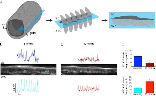
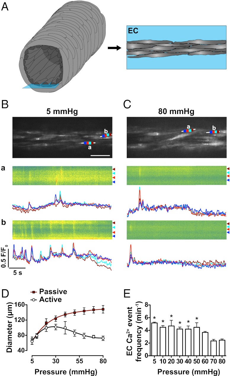
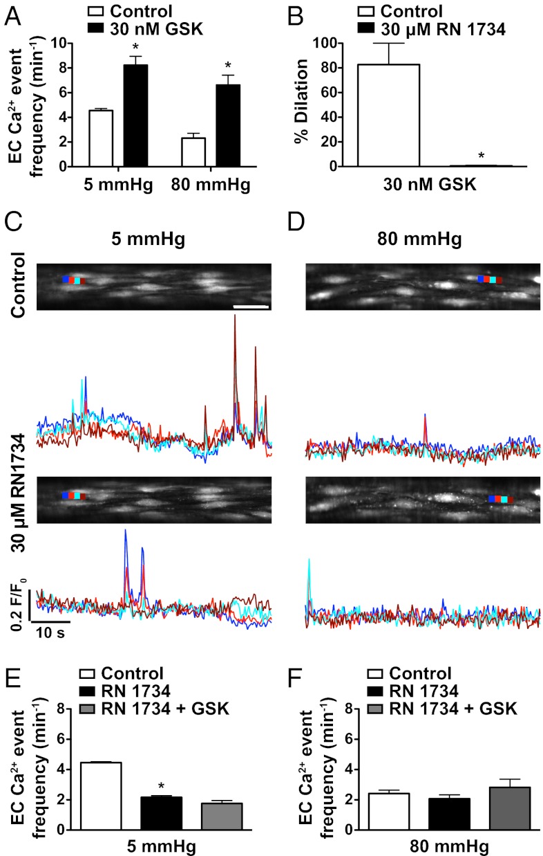
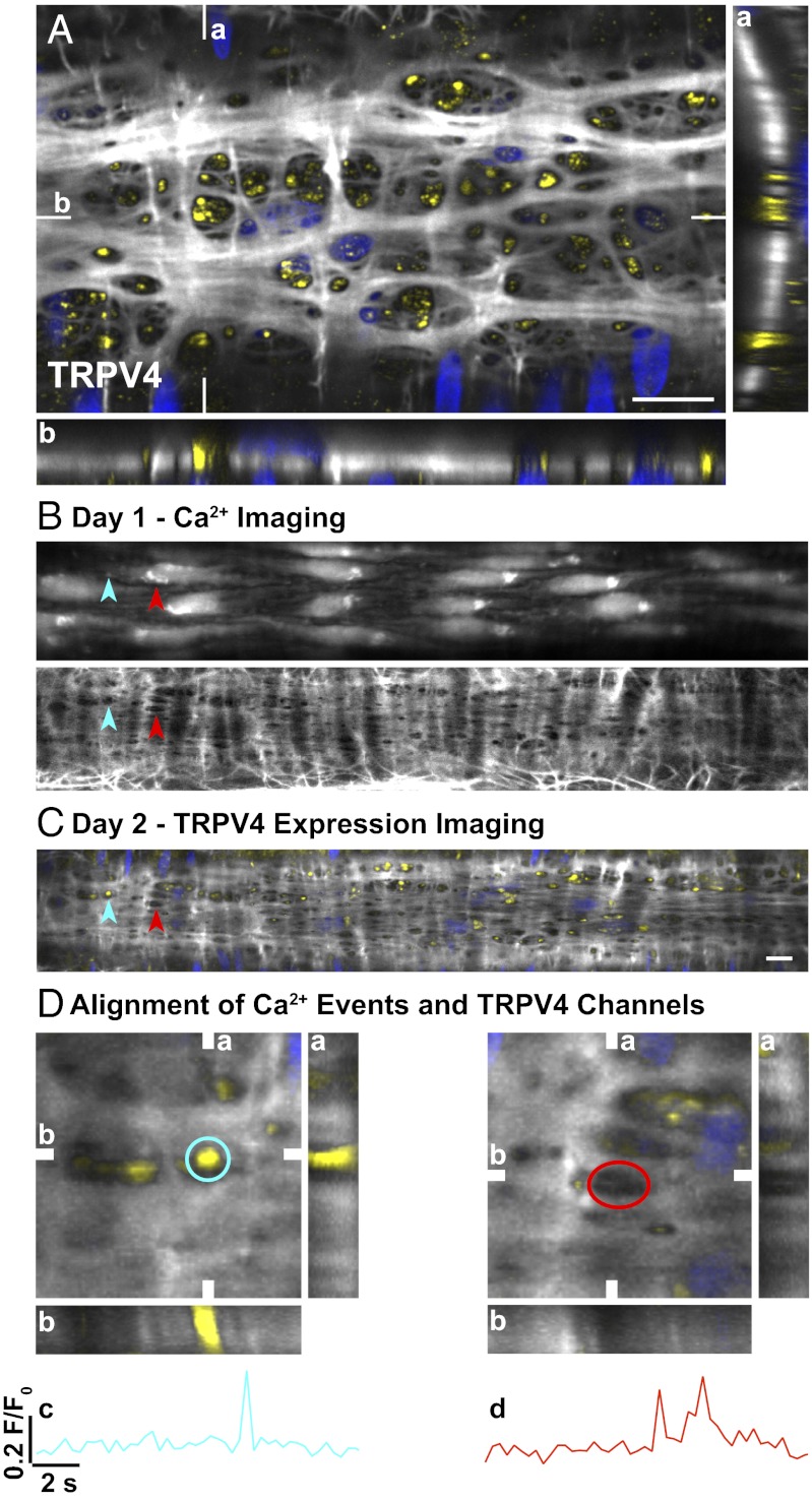
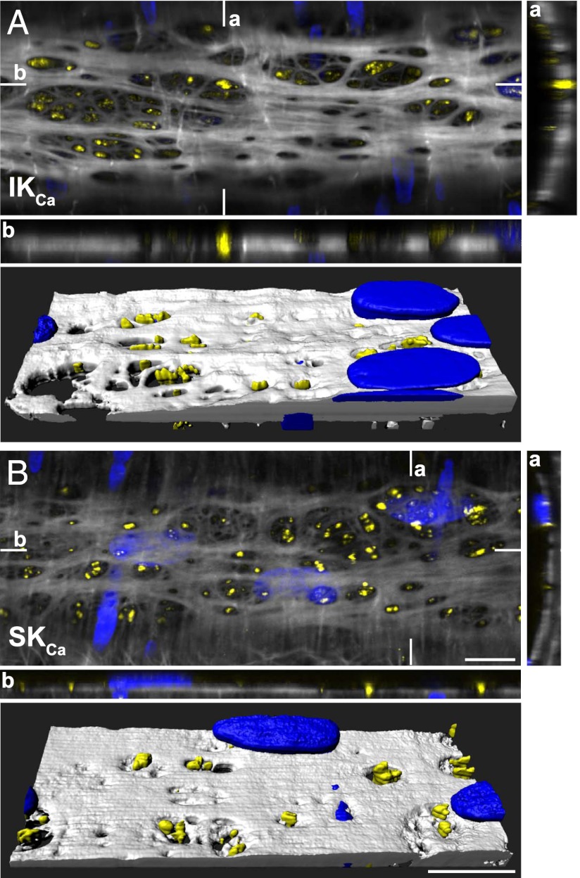
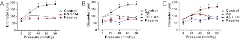
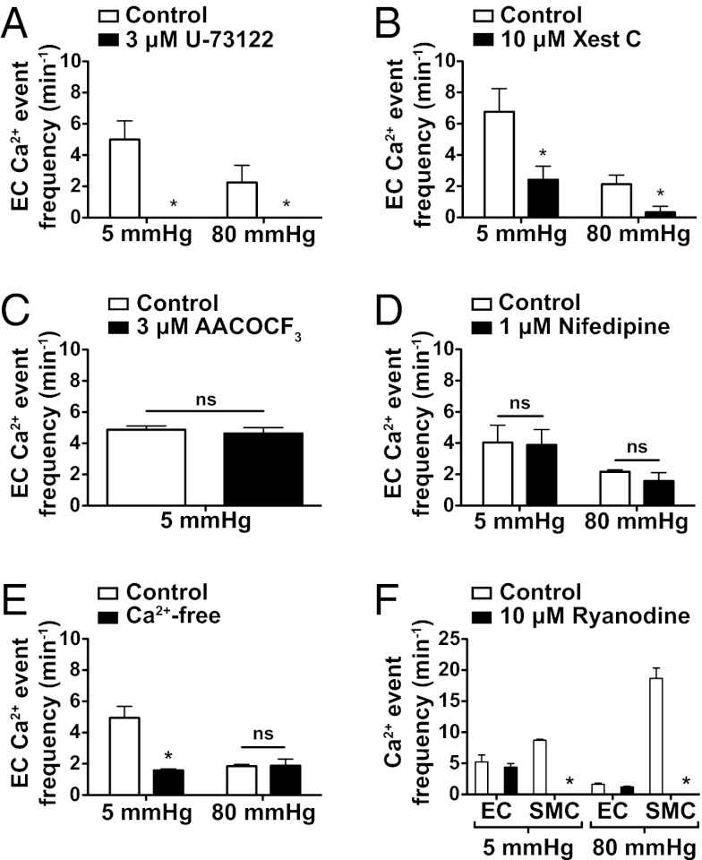
Endothelial cell (EC) Ca(2+)-activated K channels (SK(Ca) and IK(Ca) channels) generate hyperpolarization that passes to the adjacent smooth muscle cells causing vasodilation. IK(Ca) channels focused within EC projections toward the smooth muscle cells are activated by spontaneous Ca(2+) events (Ca(2+) puffs/pulsars). We now show that transient receptor potential, vanilloid 4 channels (TRPV4 channels) also cluster within this microdomain and are selectively activated at low intravascular pressure. In arterioles pressurized to 80 mmHg, ECs generated low-frequency (~2 min(-1)) inositol 1,4,5-trisphosphate receptor-based Ca(2+) events. Decreasing intraluminal pressure below 50 mmHg increased the frequency of EC Ca(2+) events twofold to threefold, an effect blocked with the TRPV4 antagonist RN1734. These discrete events represent both TRPV4-sparklet- and nonsparklet-evoked Ca(2+) increases, which on occasion led to intracellular Ca(2+) waves. The concurrent vasodilation associated with increases in Ca(2+) event frequency was inhibited, and basal myogenic tone was increased, by either RN1734 or TRAM-34 (IK(Ca) channel blocker), but not by apamin (SK(Ca) channel blocker). These data show that intraluminal pressure influences an endothelial microdomain inversely to alter Ca(2+) event frequency; at low pressures the consequence is activation of EC IK(Ca) channels and vasodilation, reducing the myogenic tone that underpins tissue blood-flow autoregulation.


Conflict of interest statement

The authors declare no conflict of interest.

Figures

Fig. 1.
Fig. 1.
Midplane imaging of spontaneous Ca2+ events in pressurized cremaster arteriolar ECs and SMCs. (A) Schematic representation of an arteriole (Left) showing the SMCs (light gray) wrapped circumferentially around the axially aligned ECs (dark gray, Center). Focusing at the midplane of an arteriole (blue box) allows both cell types to be imaged simultaneously (Right). (B and C) Midplane images of an intact arteriole pressurized to either 5 (B) or 80 (C) mmHg (ECs left to right, SMCs in cross section). Regions of interest (ROIs) shown on the image correspond to the temporal fluorescence traces from either ECs (Upper) or SMCs (Lower). (Scale bar, 10 μm; Movie S1). (D) Summary data of events recorded simultaneously in both cell types. The pressure-dependent frequency of EC Ca2+ events (Upper) changed in the opposite direction to SMCs (Lower). Note that changes in fluorescence intensity do not directly relate to amplitudes in [Ca2+]i. *P < 0.05, significantly different from 5 mmHg (n = 5). Images were acquired at ∼3 Hz.
Fig. 2.
Fig. 2.
Bottom-plane imaging of EC Ca2+ events in pressurized cremaster arterioles. (A) Imaging ECs at the lowest (Right, blue box) plane in an arteriole enables both spatial and temporal resolution of local and propagating Ca2+ events. (B and C) Confocal fluorescence images of ECs at an intraluminal pressure of 5 mmHg (B) (Movies S1 and S2) and 80 mmHg (C) in the same vessel. (Scale bar, 30 μm.) White lines indicate the site of line-scan analysis with each color (dark blue, light red, light blue, and dark red) corresponding to traces in a and b (low Ca2+, green; high Ca2+, red), raw data, with corresponding Ca2+ events from the defined subcellular regions plotted below as F/F0 over time. (D) Summary data of diameter change in pressurized arterioles, showing myogenic tone developing above 30 mmHg during step increases in intraluminal pressure; active (open circles) arterial diameter associated with an increase in the corresponding frequency of EC Ca2+ events at lower pressures (E). Note that arteriolar diameter at 5 mmHg (no tone) was effectively the same as at 80 mmHg (myogenic tone). Corresponding Ca2+-free passive diameters (red squares) for each dataset are also shown. *P < 0.05; significantly different from 80 mmHg (paired observations; n = 3). Images were acquired at ∼9 Hz.
Fig. 3.
Fig. 3.
TRPV4 channels underlie activation of EC Ca2+ events by a low-pressure-dependent mechanism. (A) The TRPV4 agonist GSK1016790A (GSK; 30 nM) increased EC Ca2+ events at both 5 and 80 mmHg. (B) In arterioles with myogenic tone at 80 mmHg, 30 nM GSK evoked 80% dilation, an effect blocked by the TRPV4 antagonist RN1734 (30 μM). (C and D) OGB-1 fluorescence reporting EC Ca2+ events in the bottom confocal plane of pressurized arterioles at an intraluminal pressure of either 5 (C) or 80 (D) mmHg before (Upper) and after (Lower) incubation with 30 μM RN1734. (Scale bar, 30 μm.) Ca2+ events in subcellular regions (color coded, dark blue, light red, light blue, and dark red) are plotted below as F/F0 changes with time. (E and F) Summary data show the effect of RN1734 against basal EC Ca2+ events at 5 (E) and 80 (F) mmHg. *P < 0.05; significantly different from control (paired observations, n = 4). In the same arterioles, RN1734 also blocked any increase in EC Ca2+ event frequency to 30 nM GSK. Images were acquired at ∼3 Hz.
Fig. 4.
Fig. 4.
Correlation between EC spontaneous Ca2+ events, holes in the IEL, and expression of TRPV4 channels in pressurized cremaster arterioles. (A) Confocal merged image of three (0.2-μm) z-axis planes of a pressurized arteriole at the level of the IEL. TRPV4 expression (yellow) localized primarily to holes through the IEL (white with nuclear staining blue). Data correlating TRPV4 expression and EC Ca2+ events were accumulated over 2 d in the same pressurized arteriole. Spontaneous EC Ca2+ events were measured, and the elastin was stained (5 mmHg; day 1). Sites of spontaneous Ca2+ activity were identified and analyzed post hoc to determine the correlation with holes in the IEL (overlay in Lower). (B) Spontaneous Ca2+ events were restricted to holes in the IEL in ∼71% of cases (n = 3 arterioles; blue and red arrowheads). (C) The arteriole was then fixed in situ, and TRPV4 expression was determined the next day (day 2). (D) EC spontaneous Ca2+ events aligned with TRPV4 expression in 58 ± 6% (n = 3) of cases (C, blue arrowhead; D, blue ROI; c, time-series plot). Spontaneous Ca2+ activity indicated by the red arrowhead in B and C aligned with a hole in the IEL but not with TRPV4 expression (D, red ROI; d, time-series plot). (A and D) Reconstructed z-stacks are shown in both the vertical (a) and horizontal (b) planes indicated on the merged image. (Scale bars, 10 μm.) Field of view in D, 23 μm2. Images were acquired at ∼3 Hz.
Fig. 5.
Fig. 5.
Association between KCa3.1 and KCa2.3 channel expression and holes through the IEL in pressurized cremaster arterioles. Confocal image from a pressurized arteriole (merged image of three 0.2-μm z-axis planes at the level of the IEL). KCa3.1 (IKCa channel; A) and KCa2.3 (SKCa channel; B) expression is indicated in yellow, IEL in white, and nuclear staining in blue. Reconstructed z-stacks are shown for the corresponding vertical (a) and horizontal (b) planes indicated on the main, merged image. Note the localization of IKCa and SKCa channels primarily within holes through the IEL, highlighted in the 3D volume reconstruction (A and B, Lower). (Scale bars, 10 μm.) Data are representative of images from three arterioles (Movie S3).
Fig. 6.
Fig. 6.
EC TRPV4 and IKCa channels suppress the development of myogenic tone in cremaster arterioles. (A and B) At low intraluminal pressure, block of either TRPV4 channels with RN1734 (30 μM, n = 4; A) or IKCa channels with TRAM-34 (TR, 1 μM, n = 3; B) significantly increased the level of myogenic tone. (C) In contrast, block of SKCa channels with apamin (Ap) did not alter the pressure/diameter profile (100 nM, n = 3). The passive diameters obtained in Ca2+-free buffer for each dataset are also shown. *P < 0.05, significantly different from control (100 μM l-NAME) at the same pressure (paired observations).
Fig. 7.
Fig. 7.
EC Ca2+ events involve IP3R signaling and Ca2+ influx but not arachidonic acid derivatives, VGCC, or ryanodine receptors. (A) EC spontaneous Ca2+ events in pressurized cremaster arterioles were completely abolished by the PLC antagonist U-73122 (3 μM) at 5 mmHg (n = 3) and 80 mmHg (n = 3). (B) IP3R blockade with xestospongin C (Xest C, 10 μM) attenuated spontaneous EC Ca2+ event frequency at both 5 mmHg (n = 3) and 80 mmHg (n = 4). (C) Block of PLA2, a liberator of arachidonic acid, with AACOCF3 (3 μM) did not alter (ns, not significant) EC Ca2+ event frequency at 5 mmHg. (D) Block of VGCC with nifedipine (1 μM) had no effect on Ca2+ event frequency at either 5 or 80 mmHg (n = 4) but prevented the development of myogenic tone. (E) Removal of extracellular Ca2+ (Ca2+-free) reduced EC activity at 5 mmHg but not at 80 mmHg (n = 3). (F) Ryanodine (10 μM) abolished asynchronous propagated SMC Ca2+ events (waves) at both 5 and 80 mmHg intraluminal pressure in cremaster arterioles (n = 3) but did not affect EC spontaneous Ca2+ activity in the same arterioles (n = 3). *P < 0.05, significantly different from control at each pressure.

References

    1. Garland CJ, Hiley CR, Dora KA. EDHF: Spreading the influence of the endothelium. Br J Pharmacol. 2011;164(3):839–852. - PMC - PubMed
    1. Bagher P, Segal SS. Regulation of blood flow in the microcirculation: role of conducted vasodilation. Acta Physiol (Oxf) 2011;202(3):271–284. - PMC - PubMed
    1. Crane GJ, Gallagher N, Dora KA, Garland CJ. Small- and intermediate-conductance calcium-activated K+ channels provide different facets of endothelium-dependent hyperpolarization in rat mesenteric artery. J Physiol. 2003;553(Pt 1):183–189. - PMC - PubMed
    1. Brähler S, et al. Genetic deficit of SK3 and IK1 channels disrupts the endothelium-derived hyperpolarizing factor vasodilator pathway and causes hypertension. Circulation. 2009;119(17):2323–2332. - PubMed
    1. Dora KA, Gallagher NT, McNeish A, Garland CJ. Modulation of endothelial cell KCa3.1 channels during endothelium-derived hyperpolarizing factor signaling in mesenteric resistance arteries. Circ Res. 2008;102(10):1247–1255. - PMC - PubMed

Publication types

LinkOut - more resources