. 2012 Sep;12(5):213-25.
doi: 10.1016/s0972-6292(16)30544-7. Epub 2012 Sep 1.

Fragmented QRS: What Is The Meaning?

Affiliations

Fragmented QRS: What Is The Meaning?

Yutaka Take et al. Indian Pacing Electrophysiol J. 2012 Sep.

Abstract

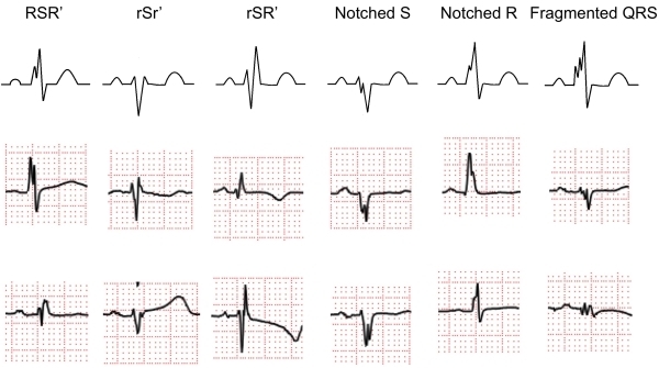
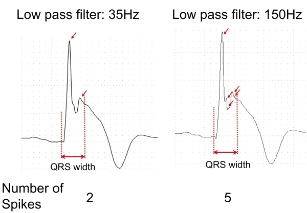
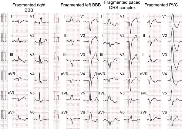
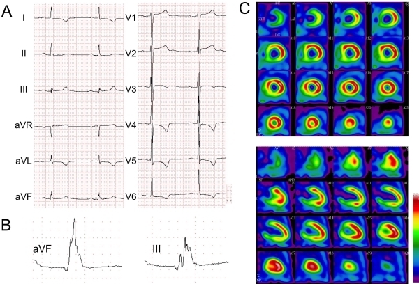
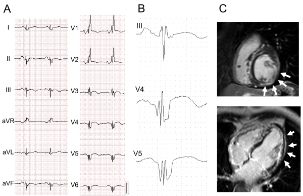
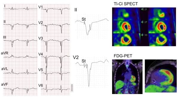
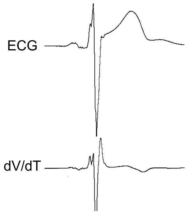
Fragmented QRS (fQRS) is a convenient marker of myocardial scar evaluated by 12-lead electrocardiogram (ECG) recording. fQRS is defined as additional spikes within the QRS complex. In patients with CAD, fQRS was associated with myocardial scar detected by single photon emission tomography and was a predictor of cardiac events. fQRS was also a predictor of mortality and arrhythmic events in patients with reduced left ventricular function. The usefulness of fQRS for detecting myocardial scar and for identifying high-risk patients has been expanded to various cardiac diseases, such as cardiac sarcoidosis, arrhythmogenic right ventricular cardiomyopathy, acute coronary syndrome, Brugada syndrome, and acquired long QT syndrome. fQRS can be applied to patients with wide QRS complexes and is associated with myocardial scar and prognosis. Myocardial scar detected by fQRS is associated with subsequent ventricular dysfunction and heart failure and is a substrate for reentrant ventricular tachyarrhythmias.

Keywords: cardiac event; cardiovascular implantable electronic device (CIED); fragmented QRS; myocardial scar.


Figures

Figure 1
Figure 1
Classification of fragmented QRS (various RSR' patterns). Fragmented QRS was defined as an additional spike of QRS complexes without bundle branch block. Various RSR' patterns are present in the mid precordial lead or inferior lead.
Figure 2
Figure 2
Effects of low-pass filter. ECG recording with a low-pass filter of 35Hz showed only 2 spikes within the QRS complex (left). Change of the cut-off frequency from 35 to 150 Hz unmasked 3 additional spikes within the QRS complex (right).
Figure 3
Figure 3
Fragmented wide QRS complex. Fragmented wide QRS complex in the bundle branch block (BBB), paced QRS complex and premature ventricular complex (PVC) have more than two notches.
Figure 4
Figure 4
Fragmented QRS in a 64-year-old patient with old myocardial infarction. A) 12-lead ECG did not have an abnormal Q wave, B) multiple R waves were present in III and aVF leads, and C) nuclear imaging (99m Tc-TF) showed a fixed inferior myocardial perfusion defect.
Figure 5
Figure 5
Fragmented QRS in a patient with dilated cardiomyopathy. ECG and images from a 74-year-old patient with left ventricular dysfunction (ejection fraction: 32%). The patient was diagnosed as having non-ischemic dilated cardiomyopathy. A) 12-lead ECG showed right bundle branch block, B) fQRS (various RSR' patterns) was present in left lateral and inferior leads, and C) delayed enhancement in Ga-MRI was present in the inferolateral resion (white arrows).
Figure 6
Figure 6
Fragmented QRS in a 52-year-old patient with cardiac sarcoidosis. A) 12-lead ECG showed a wide QRS complex by right ventricular apex pacing, B) fQRS was present in inferior and mid-precordial leads, C) Thalium-201 myocardial perfusion imaging showed an anterolateral-infero perfusion defect. 18F-fluoro-2-deoxyglucose positron emission tomography (18F-FDG-PET) showed FDG accumulation at the same site of the perfusion defect.
Figure 7
Figure 7
New analysis of fragmented QRS using the first derivation of voltage by time (dV/dT) of the QRS complex. Spikes and notches within the QRS complex were clearly represented as positive peaks in the dV/dT analysis. ECG and dV/dT of the QRS complex were recorded by FX-7524 of Fukuda Denshi Co. Ltd and analyzed by original software.

References

    1. Surawicz B, et al. Chou's electrocardiography in clinical practice. Philadelphia: Elsevier; 2008.
    1. Varriale P, et al. The RSR' complex not related to right bundle branch block: diagnostic value as a sign of myocardial infarction scar. Am Heart J . 1992;123:369. - PubMed
    1. Das MK, et al. Significance of a fragmented QRS complex versus a Q wave in patients with coronary artery disease. Circulation . 2006;113:2495. - PubMed
    1. Das MK, et al. Fragmented wide QRS on a 12-lead ECG: a sign of myocardial scar and poor prognosis. Circ Arrhythm Electrophysiol . 2008;1:258. - PubMed
    1. Das MK, et al. Fragmented QRS on a 12-lead ECG: a predictor of mortality and cardiac events in patients with coronary artery disease. Heart Rhythm . 2007;4:1385. - PubMed

LinkOut - more resources