

MOSAIC: a multiscale model of osteogenesis and sprouting angiogenesis with lateral inhibition of endothelial cells
- PMID: 23071433
- PMCID: PMC3469420
- DOI: 10.1371/journal.pcbi.1002724
MOSAIC: a multiscale model of osteogenesis and sprouting angiogenesis with lateral inhibition of endothelial cells
Erratum in
- PLoS Comput Biol. 2013 Mar;9(3). doi: 10.1371/annotation/38264a13-d4b5-49cd-b54e-47330bb19fe9 doi: 10.1371/annotation/38264a13-d4b5-49cd-b54e-47330bb19fe9
Abstract
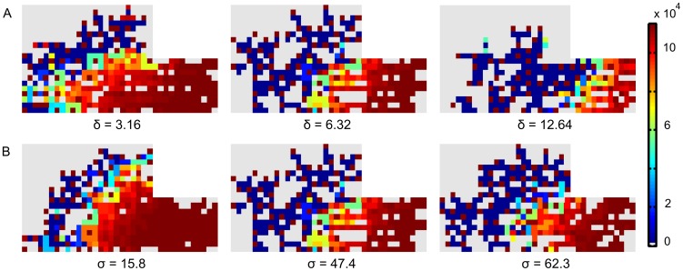
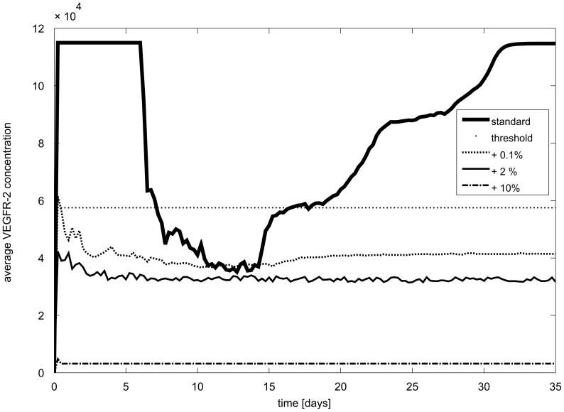
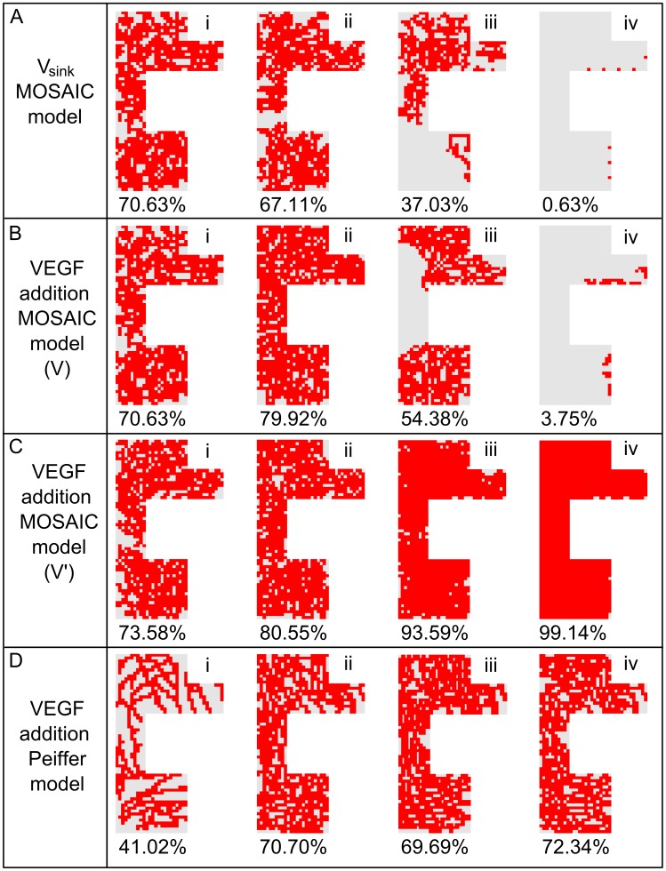
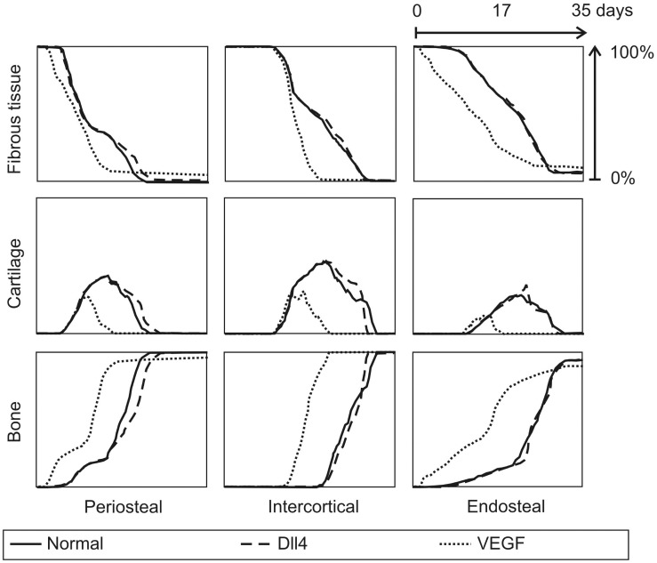
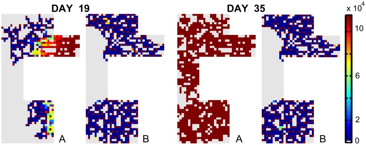
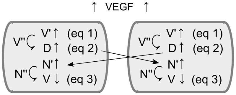
The healing of a fracture depends largely on the development of a new blood vessel network (angiogenesis) in the callus. During angiogenesis tip cells lead the developing sprout in response to extracellular signals, amongst which vascular endothelial growth factor (VEGF) is critical. In order to ensure a correct development of the vasculature, the balance between stalk and tip cell phenotypes must be tightly controlled, which is primarily achieved by the Dll4-Notch1 signaling pathway. This study presents a novel multiscale model of osteogenesis and sprouting angiogenesis, incorporating lateral inhibition of endothelial cells (further denoted MOSAIC model) through Dll4-Notch1 signaling, and applies it to fracture healing. The MOSAIC model correctly predicted the bone regeneration process and recapitulated many experimentally observed aspects of tip cell selection: the salt and pepper pattern seen for cell fates, an increased tip cell density due to the loss of Dll4 and an excessive number of tip cells in high VEGF environments. When VEGF concentration was even further increased, the MOSAIC model predicted the absence of a vascular network and fracture healing, thereby leading to a non-union, which is a direct consequence of the mutual inhibition of neighboring cells through Dll4-Notch1 signaling. This result was not retrieved for a more phenomenological model that only considers extracellular signals for tip cell migration, which illustrates the importance of implementing the actual signaling pathway rather than phenomenological rules. Finally, the MOSAIC model demonstrated the importance of a proper criterion for tip cell selection and the need for experimental data to further explore this. In conclusion, this study demonstrates that the MOSAIC model creates enhanced capabilities for investigating the influence of molecular mechanisms on angiogenesis and its relation to bone formation in a more mechanistic way and across different time and spatial scales.
Conflict of interest statement
The authors have declared that no competing interests exist.
Figures
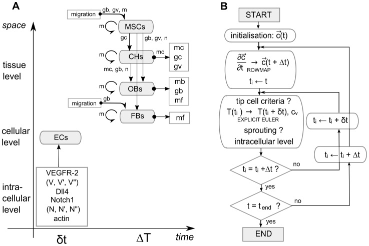
 : vector of the continuous variables, t: time, δt: time step of the inner loop, Δt: time step of the outer loop, cv: endothelial cells).
References
-
- Taguchi K, Ogawa R, Migita M, Hanawa H, Ito H, et al. (2005) The role of bone marrow-derived cells in bone fracture repair in a green fluorescent protein chimeric mouse model. Biochem Biophys Res Commun 331: 31–36. - PubMed
-
- Carmeliet P, De Smet F, Loges S, Mazzone M (2009) Branching morphogenesis and antiangiogenesis candidates: tip cells lead the way. Nat Rev Clin Oncol 6: 315–326. - PubMed
-
- Bentley K, Gerhardt H, Bates PA (2008) Agent-based simulation of notch-mediated tip cell selection in angiogenic sprout initialisation. J Theor Biol 250: 25–36 S0022-5193(07)00443-2 [pii];10.1016/j.jtbi.2007.09.015 [doi]. - PubMed
-
- De Smet F, Segura I, De Bock K, Hohensinner PJ, Carmeliet P (2009) Mechanisms of Vessel Branching Filopodia on Endothelial Tip Cells Lead the Way. Arterioscler Thromb Vasc Biol 29: 639–649. - PubMed
-
- Hellstrom M, Phng LK, Hofmann JJ, Wallgard E, Coultas L, et al. (2007) Dll4 signalling through Notch1 regulates formation of tip cells during angiogenesis. Nature 445: 776–780. - PubMed
