. 2012;8(10):e1002959.
doi: 10.1371/journal.ppat.1002959. Epub 2012 Oct 11.

Human cytomegalovirus infection dysregulates the canonical Wnt/β-catenin signaling pathway

Affiliations

Human cytomegalovirus infection dysregulates the canonical Wnt/β-catenin signaling pathway

Magdalena Angelova et al. PLoS Pathog. 2012.

Abstract

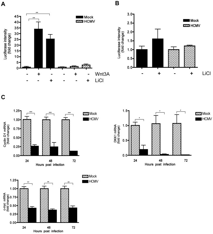
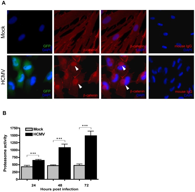
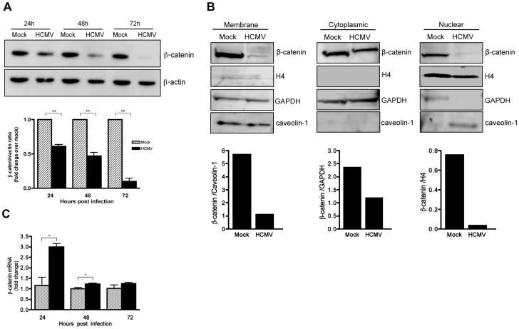
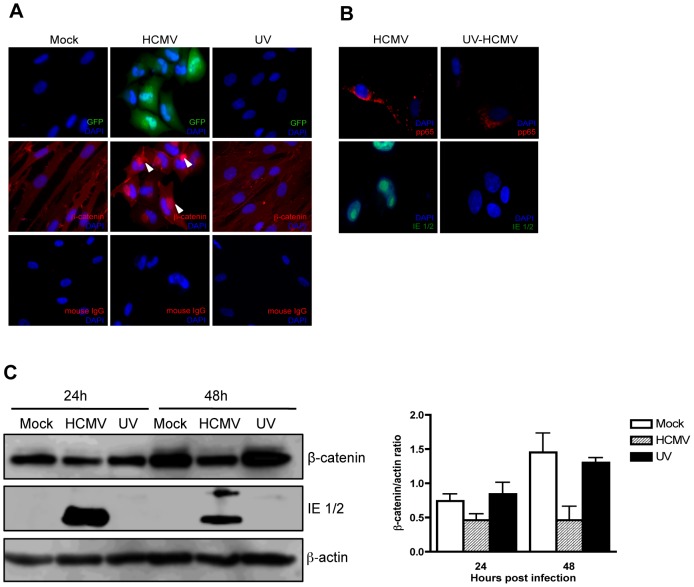
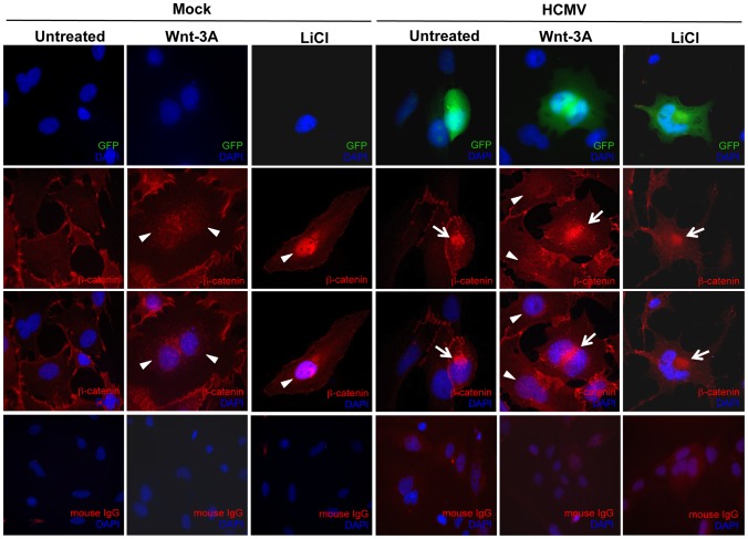
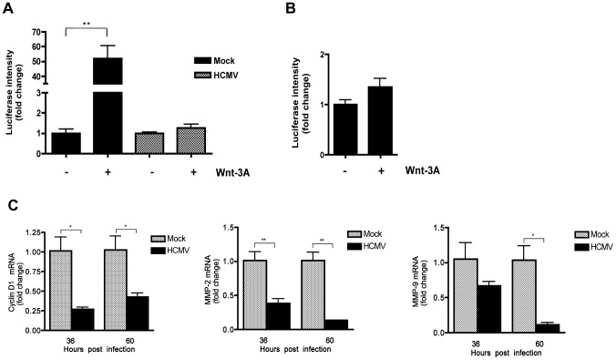
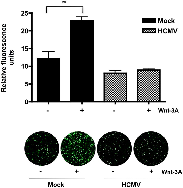
Human Cytomegalovirus (HCMV) is a ubiquitous herpesvirus that currently infects a large percentage of the world population. Although usually asymptomatic in healthy individuals, HCMV infection during pregnancy may cause spontaneous abortions, premature delivery, or permanent neurological disabilities in infants infected in utero. During infection, the virus exerts control over a multitude of host signaling pathways. Wnt/β-catenin signaling, an essential pathway involved in cell cycle control, differentiation, embryonic development, placentation and metastasis, is frequently dysregulated by viruses. How HCMV infection affects this critical pathway is not currently known. In this study, we demonstrate that HCMV dysregulates Wnt/β-catenin signaling in dermal fibroblasts and human placental extravillous trophoblasts. Infection inhibits Wnt-induced transcriptional activity of β-catenin and expression of β-catenin target genes in these cells. HCMV infection leads to β-catenin protein accumulation in a discrete juxtanuclear region. Levels of β-catenin in membrane-associated and cytosolic pools, as well as nuclear β-catenin, are reduced after infection; while transcription of the β-catenin gene is unchanged, suggesting enhanced degradation. Given the critical role of Wnt/β-catenin signaling in cellular processes, these findings represent a novel and important mechanism whereby HCMV disrupts normal cellular function.


Conflict of interest statement

The authors have declared that no competing interests exist.

Figures

Figure 1
Figure 1. Inhibition of β-catenin transcriptional activity by HCMV.
(A) HFFs were co-transfected with TOPflash and pRL-TK plasmids and 12 hr later infected with HCMV-TR (MOI of 1) or mock-infected. Forty eight hr after infection, the cells were stimulated with either 150 ng/mL Wnt-3A, 20 mM LiCl or PBS control for an additional 12 hr. Luciferase expression in cell lysates was measured and normalized to Renilla luciferase activity. Data are presented as the mean of results from two independent experiments each performed with triplicate transfections. **p<0.01. (B) HFFs were transfected with FOPflash plasmid (as negative control) and pRL-TK control plasmid and mock- or TR-infected, then stimulated with LiCl and analyzed as described in (A). (C) Cyclin D1, DKK1 and c-myc mRNA levels in HFFs were analyzed from triplicate infections by qRT-PCR at 24, 48 and 72 hr after infection with HCMV-TR (MOI of 1). Each sample was analyzed in triplicate and normalized to the level of 36B4. The data are presented as fold change (mean +/− SEM) relative to the mock-infected sample at each corresponding time point. *p<0.05; **p<0.01; ***p<0.001.
Figure 2
Figure 2. β-catenin aggregates at a central juxtanuclear location in HCMV-infected HFFs.
(A) HFFs were seeded on glass coverslips and infected with Towne-GFP (MOI of 1–2) for 48 hr. Cells were stained with mouse monoclonal antibody to β-catenin followed by AlexaFluor 555-conjugated anti-mouse IgG. Nuclei were counterstained with DAPI. Normal mouse IgG was used as an isotype control. GFP positive cells represent infected cells. Arrowheads indicate aggregation of β-catenin in infected cells. (B) Cell extracts collected from four separate wells of HHFs at 24, 48 or 72 hr after infection with HCMV-TR (MOI of 1–2) were analyzed for the chymotrypsin-like activity of the 26S proteasome by addition of a fluorogenic peptide substrate (Suc-LLVY-AMC). Proteasome activity is expressed as relative fluorescence units and reported as the mean ± SEM (n = 4) and is representative of two independent experiments. ***p<0.001.
Figure 3
Figure 3. HCMV induces degradation of β-catenin.
(A) Lysates were collected from HFFs infected with HCMV-TR (MOI of 1–2) at the indicated times post infection and analyzed for β-catenin expression by Western blot. β-actin served as a loading control. β-catenin protein levels were quantitated by densitometric analysis using ImageJ software (n = 3) and normalized to mock-infected cells at each time point, which was set to a value of 1.0. Data are presented as the mean ± SEM of 3 independent experiments. **p<0.01 (B) Membrane, cytoplasmic and nuclear protein-enriched cell fractions were prepared from HFFs 48 hr after infection with HCMV-TR (MOI of 1–2), and analyzed for β-catenin expression by Western blot. Caveolin-1, GAPDH, and histone H4 served as loading controls for the membrane, cytoplasmic and nuclear fractions, respectively. β-catenin protein levels were quantitated by densitometric analysis using ImageJ software. (C) β-catenin mRNA expression in HCMV-TR-infected (MOI of 1–2) or mock-infected HFFs at 24, 48 and 72 hr post infection were analyzed by qRT-PCR. Relative β-catenin mRNA levels in virus-infected samples at each timepoint were normalized to GAPDH mRNA levels and expressed as fold change relative to the corresponding uninfected control. Data are presented as mean +/− SEM of 3 independent experiments. *p<0.05.
Figure 4
Figure 4. Effect of UV-inactivated HCMV on β-catenin degradation.
(A) HFFs grown on chamber slides were infected with Towne-GFP (MOI of 1–2), UV-irradiated Towne-GFP or mock-infected and fixed at 48 hr post infection. The cells were stained with mouse anti-β-catenin antibody followed by AlexaFluor 555-conjugated anti-mouse IgG. Nuclei were counterstained with DAPI. Mouse IgG was used as an isotype control. GFP positive cells represent infected cells. Arrowheads indicate aggregation of β-catenin in infected cells. (B) Immunofuorescence detection of the viral tegument protein pp65 (red) and immediate early proteins (green) in HFFs infected (MOI of 1–2) with HCMV-TR and UV-irradiated HCMV-TR at 6 hr (pp65) and 24 hr (IE1/2) post infection. (C) HFFs were infected with HCMV-TR, UV-irradiated HCMV-TR or mock-infected (MOI of 1–2). Protein lysates were collected at 24 and 48 hr post infection and analyzed for β-catenin and HCMV IE 1/2 expression by Western blot followed by quantitative densitometric analysis using ImageJ software. β-actin served as a loading control. Data are presented as mean +/− SEM of 2 independent experiments.
Figure 5
Figure 5. HCMV infection inhibits β-catenin nuclear translocation in EVTs.
SGHPL-4 cells were seeded on glass coverslips and infected with Towne-GFP (MOI of 1–2) or mock-infected for 48 hr. Cells were then treated with 150 ng/ml Wnt-3A or 20 mM LiCl for 6 hr followed by immunostaining for β-catenin using a mouse anti-β catenin antibody, followed by a goat anti-mouse secondary IgG conjugated to AlexaFluor 555. Nuclei were counterstained with DAPI. Mouse IgG was used as an isotype control. GFP positive cells represent infected cells. Arrowheads point to nuclear accumulation of β-catenin. Arrows indicate aggregation of β-catenin in infected cells.
Figure 6
Figure 6. HCMV infection inhibits Wnt/β-catenin transcriptional activity in EVTs in response to Wnt stimulation.
(A) SGHPL-4 cells were co-transfected with TOPflash and pRL-TK plasmids and 6 hr later infected with Towne-GFP (MOI of 1–2) or mock-infected. 48 hr after infection, the cells were stimulated with either 150 ng/mL Wnt-3A or PBS control for an additional 12 hr. Luciferase expression in cell lysates was measured and normalized to Renilla luciferase activity. Data are presented as the mean ± SEM of results from three independent experiments each performed with duplicate transfections (n = 6). **p<0.01. (B) FOPflash (C) Cyclin D1, MMP-2 and MMP-9 mRNA levels in SGHPL-4 cells 36 and 60 hr after infection with HCMV-TR (MOI of 1–2) or mock-infection were analyzed by qRT-PCR in triplicate. mRNA levels were normalized to GAPDH expression. The data are presented as fold change (mean +/− SEM) relative to the mock-infected sample at each timepoint. *p<0.05; **p<0.01.
Figure 7
Figure 7. HCMV inhibits Wnt-3A-induced migration of SGHPL-4 EVTs.
Migration was assayed using BD FluoroBlok transwell inserts. SGHPL-4 cells were infected with Towne-GFP (MOI of 1–2) or mock-infected for 48 hr prior to the assay. Equal numbers of cells were loaded into the inserts and 150 ng/ml of Wnt-3A or PBS were added to both the upper and lower chambers of the transwell system as indicated. Each condition was performed in triplicate. Migrated wells were stained with calcein AM and visualized with a fluorescent microscope. Average fluorescence intensity was determined by capturing three random fields from each well and measuring their fluorescence intensity using ImageJ software. Data are represented as the mean ± SEM (n = 9). **p<0.001.

References

    1. Steininger C (2007) Clinical relevance of cytomegalovirus infection in patients with disorders of the immune system. Clin Microbiol Infect 13: 953–963. - PubMed
    1. Cannon MJ (2009) Congenital cytomegalovirus (CMV) epidemiology and awareness. J Clin Virol 46 (Suppl 4) S6–10. - PubMed
    1. Trincado DE, Rawlinson WD (2001) Congenital and perinatal infections with cytomegalovirus. J Paediatr Child Health 37: 187–192. - PubMed
    1. Demmler GJ (1996) Congenital cytomegalovirus infection and disease. Adv Pediatr Infect Dis 11: 135–162. - PubMed
    1. Nigro G, Adler SP (2011) Cytomegalovirus infections during pregnancy. Curr Opin Obstet Gynecol 23: 123–128. - PubMed

Publication types