. 2012;8(10):e1002965.
doi: 10.1371/journal.pgen.1002965. Epub 2012 Oct 11.

Phosphatase-dead myotubularin ameliorates X-linked centronuclear myopathy phenotypes in mice

Affiliations

Phosphatase-dead myotubularin ameliorates X-linked centronuclear myopathy phenotypes in mice

Leonela Amoasii et al. PLoS Genet. 2012.

Abstract

Myotubularin MTM1 is a phosphoinositide (PPIn) 3-phosphatase mutated in X-linked centronuclear myopathy (XLCNM; myotubular myopathy). We investigated the involvement of MTM1 enzymatic activity on XLCNM phenotypes. Exogenous expression of human MTM1 in yeast resulted in vacuolar enlargement, as a consequence of its phosphatase activity. Expression of mutants from patients with different clinical progression and determination of PtdIns3P and PtdIns5P cellular levels confirmed the link between vacuolar morphology and MTM1 phosphatase activity, and showed that some disease mutants retain phosphatase activity. Viral gene transfer of phosphatase-dead myotubularin mutants (MTM1(C375S) and MTM1(S376N)) significantly improved most histological signs of XLCNM displayed by a Mtm1-null mouse, at similar levels as wild-type MTM1. Moreover, the MTM1(C375S) mutant improved muscle performance and restored the localization of nuclei, triad alignment, and the desmin intermediate filament network, while it did not normalize PtdIns3P levels, supporting phosphatase-independent roles of MTM1 in maintaining normal muscle performance and organelle positioning in skeletal muscle. Among the different XLCNM signs investigated, we identified only triad shape and fiber size distribution as being partially dependent on MTM1 phosphatase activity. In conclusion, this work uncovers MTM1 roles in the structural organization of muscle fibers that are independent of its enzymatic activity. This underlines that removal of enzymes should be used with care to conclude on the physiological importance of their activity.


Conflict of interest statement

The authors have declared that no competing interests exist.

Figures

Figure 1
Figure 1. Human myotubularin expression in yeast S. cerevisiae.
(A) Representation of the MTM1 protein with its domains, the position of the mutations analyzed and the severity of the resulting myopathy phenotype. MTM1 displays different domains, PH-GRAM (pleckstrin homology-glucosyltransferase, Rab-like GTPase activator and myotubularin), RID (Rac-induced recruitment domain), catalytic phosphatase domain and SID (SET-protein interaction domain). (B) Anti-MTM1 Western-blot on yeast protein extracts. The MTM1 gene was placed under the control of the tetracycline-repressible tetO promoter in the low copy number centromeric plasmid (CEN, pVV204, 1 to 3 copies per cell). In the high copy number 2 µ plasmid (2 µ, pVV200, 20 to 50 copies per cell), the MTM1 gene was under the control of the strong yeast PGK1 promoter. These different plasmids were transformed into the ymr1Δ yeast mutant. Protein extracts of ymr1Δ cells transformed with pVV204 (CEN) or pVV200 (2 µ) empty plasmids (Control) or bearing the different MTM1 forms were analyzed by Western-blot. MTM1 production was detected with the mouse monoclonal 1G6 anti-MTM1 antibody. The different human MTM1 proteins were produced at the expected molecular weight (70 kDa) as compared to the ymr1Δ cells transformed with empty control plasmids that displayed no signal. Protein loading was evaluated by immunodetection of the yeast endogenous 3-phosphoglycerate kinase Pgk1 protein.
Figure 2
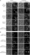
Figure 2. Yeast vacuolar phenotype analysis upon MTM1 expression.
The wild-type and fab1Δ cells were used as controls. The different yeast cells transformed or not with pVV200 (2 µ, overexpression) plasmids bearing or not wild-type or mutants MTM1 were grown to exponential phase in selective SC-trp medium and the vacuoles were stained by FM4-64. Cell were labeled with FM4-64 for 15 min at 25°C in YPD and washed in once in SC-trp. Cells were then observed in selective medium by fluorescence microscopy with DIC (Nomarski) and TRITC (FM4-64) filters. Osmotic shock was induced by addition of NaCl (final concentration 0.9 M) to FM4-64 stained cultures, and cells were observed after 10 min incubation. (A) Wild-type, fab1Δ and ymr1Δ yeast cells transformed with pVV200 empty plasmid or bearing wild-type MTM1 or the phosphatase-dead MTM1C375S mutant. Inset shows an increased magnification of a representative cell to illustrate the vacuolar phenotype upon osmotic shock. (B) ymr1Δ cells transformed with pVV200 plasmid bearing the different MTM1 mutants responsible for XLCNM myopathy.
Figure 3
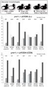
Figure 3. Vacuolar morphologies quantification in yeast cells producing MTM1.
The ymr1Δ cells expressing either wild-type MTM1 or the different MTM1 mutants from either pVV200 (2 µ, overexpression) or pVV204 (CEN, expression) plasmid were analyzed. For each strain, 300 to 600 cells were observed by microscopy (DIC and FM4-64) and sorted into one of the three categories: unilobar large or giant (in white), small one or two lobes (in grey) and more than two lobes or fragmented (in black) vacuoles. The main vacuolar phenotype of the non-transformed ymr1Δ mutant cells is fragmented vacuoles with more than two lobes. Histograms are the mean of three independent experiments and show the proportion of each category in the different transformed yeast cells.
Figure 4
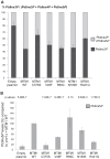
Figure 4. Determination of different phosphoinositides (PPIn) levels upon expression of MTM1 wild-type or mutant proteins.
(A) Quantification of PtdIns3P cellular levels of ymr1Δ cells expressing wild-type MTM1 or the different mutants from pVV204 (CEN). Strains were grown to early log phase in selective medium, labeled with 32P and lipids were extracted and prepared for HPLC analysis. Based on HPLC chromatograms the peak area corresponding to each PPIn species was determined and results were expressed as the percentage of 32P-PtdIns3P compared to the total labeled 32P-PtdInsP. Results are represented as the mean of at least two independent experiments shown with standard deviations. (B) Quantitative analysis of PtdIns5P produced in ymr1Δ cells expressing wild-type or mutants MTM1. Strains were grown to log phase in selective medium, lipid were extracted, separated by TLC and spots corresponding to mono-phosphorylated PPIn were scrapped off and subjected to phosphorylation by PtdIns5P 4-kinase type IIα in presence of [γ-32P]-ATP. This kinase is specific for PtdIns5P and produces PtdIns(4,5)P 2. The 32P-labeled PtdIns(4,5)P 2 generated from this in vitro kinase reaction was further analyzed by TLC and radioactivity was quantified in a scintillation counter. The total amount of PtdIns5P (pmol) in each sample was determined by comparison with a calibration curve made from diC16-PtdIns5P and normalized to the number of yeast cells. Graph represents PtdIns5P as a percentage of production compared to the wild type myotubularin MTM1 (n = 3 to 4 experiments). The p-value for each construct was evaluated versus the wild type MTM1 and is indicated at the top of the graph. A p-value of less than 0.05 indicates that the difference in the two PtdIns5P percentages is statistically significant. The p-value was 0.0021 for MTM1V49F versus MTM1C375S and 0.0036 for MTM1V49F versus MTM1R421Q.
Figure 5
Figure 5. Amelioration of muscle atrophy and fiber hypotrophy in a murine model of centronuclear myopathy by AAV–mediated expression of two phosphatase-inactive myotubularin mutants MTM1C375S and MTM1S376N.
(A) Skeletal muscle protein lysates 4 weeks post-injection were immunoblotted for MTM1 and GAPDH levels in WT and Mtm1 KO mice injected with empty AAV, Mtm1 KO injected with AAV-Mtm1-WT, AAV-Mtm1-CS and AAV- Mtm1-SN. (B) Hematoxylin and eosin (HE, left panels, magnification ×400) and succinate dehydrogenase (SDH, right panels, ×400) staining of TA cross-sections from WT mice injected with empty AAV, Mtm1 KO injected with empty AAV, AAV-Mtm1-WT, AAV-Mtm1-CS and AAV-Mtm1-SN mice 4 weeks PI. Mtm1 KO muscle sections show the presence of very small myofibers and internalized nuclei. Mitochondrial oxidative staining is abnormally accumulated at the centre of fibers. Note the recovery of oxidative reactivity pattern in myotubularin-expressing Mtm1 KO muscles. Scale bar: 50 µm. (C) Graph represents TA weight as a percentage of total body weight (n = 6 mice). P values<0.01 for WT injected with AAV versus KO injected with AAV, AAV-Mtm1-WT, AAV-Mtm1-CS and AAV-Mtm1-SN. P values<0.001 for KO injected with AAV versus AAV-Mtm1-WT, AAV-Mtm1-CS and AAV-Mtm1-SN. (D) Percentage of muscle fibers with internalized nuclei after AAV, AAV-Mtm1-WT, AAV-Mtm1-CS and AAV-Mtm1-SN injection of Mtm1 KO mice. Nuclei were considered as internalized if not in contact with the sarcolemma. The number of fibers with internal nuclei is increased in Mtm1 KO tibialis anterior (TA) muscle and significantly and equally reduced after injection with AAV-Mtm1-WT or AAV-Mtm1-CS or AAV-Mtm1-SN (n = 550). P values<0.009 for WT injected with AAV versus KO injected with AAV, AAV-Mtm1-WT, AAV-Mtm1-CS and AAV-Mtm1-SN. P values p<0.0009 for KO injected with AAV versus AAV-Mtm1-WT, AAV-Mtm1-CS and AAV-Mtm1-SN. (E) Transverse muscle sections were analyzed for fiber area. Fiber size is grouped into 200 µm2 intervals, and represented as the percentage of total fibers in each group (n = 1,000 for 15 mice for AAV-Mtm1-WT, AAV-Mtm1-CS and 6 mice for AAV-Mtm1-SN group).
Figure 6
Figure 6. The phosphatase-dead C375S myotubularin mutant improves muscle force in Mtm1 KO mice.
The specific maximal force (sP0) of WT muscle injected with AAV and Mtm1 KO TA muscle injected with AAV, AAV-Mtm1-WT and AAV-Mtm1-CS. The sP0 represents the absolute maximal force related to muscle weight (n = 6 mice, *P<0.001).
Figure 7
Figure 7. The phosphatase-dead C375S myotubularin mutant injection in Mtm1-KO muscle restores normal desmin expression and localization.
(A) Ectopic expression of MTM1 transgene in Mtm1-KO muscle restored normal desmin localization in muscle. Arrowheads indicate aggregates of desmin in Mtm1-KO muscle injected with AAV. Scale bars: 10 µm. (B) The phosphatase-dead C375S myotubularin mutant expression in Mtm1-KO muscle restored normal desmin expression level in soluble and insoluble fraction. (C) Skeletal muscle protein lysates 4 weeks post-injection were immunoblotted for MTM1. (D) Quantification of relative desmin expression level in soluble compared to insoluble fraction. Data correlated from 2 independent experiments (n = 3 mice per group). *P≤0.05. (E) Quantification of relative expression of MTM1 in WT and Mtm1 KO mice injected with empty AAV, Mtm1 KO injected with AAV-Mtm1-WT and AAV-Mtm1-CS. (F) Microsome fractions from Mtm1 KO muscles injected with AAV-Mtm1-WT and AAV-Mtm1-CS and from wild-type muscles were prepared and immunoblotted for MTM1 to compare localization of MTM1-WT and MTM1-C375S in the membrane fraction of the muscle. Microsome fractions were immunoblotted with antibodies detecting membrane proteins (SERCA1 and Sarcoglycan), cytoplasmic protein (β-tubulin) and nuclear protein (TATA-box binding protein (TBP)).
Figure 8
Figure 8. Improvement of triad abnormalities present in Mtm1-deficient muscles with AAV-Mtm1-WT and AAV-Mtm1-CS.
(A) Sarcomere and triad (arrowheads) organization in wild-type muscle, Mtm1 KO muscle, Mtm1 KO muscles injected with AAV-Mtm1-WT and AAV-Mtm1-CS at 2 different magnifications. Muscles from Mtm1 KO demonstrate a severe disorganization of the muscle fiber with lack of recognizable triads within the sarcomere structure. (B) Quantification of the presence of triads in the muscle fibers. The graph represents the ratio between the number of triads observed in each longitudinal section divided by the total number of sarcomeres present in the section. (C) Triads shape in wild-type muscle, and Mtm1 KO muscles injected with AAV-Mtm1-WT and AAV-Mtm1-CS.
Figure 9
Figure 9. MTM1-WT but not MTM1-C375S normalizes PtdIns3P levels in the injected muscles.
Lipids were extracted from wild-type, Mtm1 KO, and Mtm1-KO injected with AAV-Mtm1-WT and AAV-Mtm1-CS tibialis anterior muscles and PtdIns3P levels were quantified. The results were presented as the levels of PtdIns3P to the total phospholipids. The graphs represent the mean of two independent experiments shown with the standard deviation. *p<0.05.
Figure 10
Figure 10. XLCNM phenotypes that are ameliorated by the MTM1-C375S phosphatase-dead mutant to a similar extend as with the wild-type MTM1, in the Mtm1 KO muscle.
Phosphate-independent and phosphatase-dependent functions are underlined.

References

    1. Laporte J, Hu LJ, Kretz C, Mandel JL, Kioschis P, et al. (1996) A gene mutated in X-linked myotubular myopathy defines a new putative tyrosine phosphatase family conserved in yeast. Nat Genet 13: 175–182. - PubMed
    1. Jungbluth H, Wallgren-Pettersson C, Laporte J (2008) Centronuclear (myotubular) myopathy. Orphanet J Rare Dis 3: 26. - PMC - PubMed
    1. Biancalana V, Caron O, Gallati S, Baas F, Kress W, et al. (2003) Characterisation of mutations in 77 patients with X-linked myotubular myopathy, including a family with a very mild phenotype. Hum Genet 112: 135–142. - PubMed
    1. Buj-Bello A, Laugel V, Messaddeq N, Zahreddine H, Laporte J, et al. (2002) The lipid phosphatase myotubularin is essential for skeletal muscle maintenance but not for myogenesis in mice. Proc Natl Acad Sci U S A 99: 15060–15065. - PMC - PubMed
    1. Laporte J, Biancalana V, Tanner SM, Kress W, Schneider V, et al. (2000) MTM1 mutations in X-linked myotubular myopathy. Hum Mutat 15: 393–409. - PubMed

Publication types

MeSH terms

Substances

LinkOut - more resources